|
|
1.INTRODUCTIONOptics has always been a fascinating domain to approach, as light is a major component of human life. Thus, light and vision to begin with, then instrumentation and nowadays Photonics with so many applications hardly to present in this limited space represent fields that no scientist can miss. The path towards an optics education and why not a career, has at least three possible starting points: Physics, Electrical and Mechanical Engineering. Of course, these are not exclusive, as Biology and Medicine are also now very much into this equation, as we shall see in this presentation as well. Fine (or precision) mechanics has been particularly dedicated to optics education & research in Romania. While in the capital city of Bucharest it started in what is now UPB – the “Politehnica” University of Bucharest, in the Western (industrialized) part of the country this developement was mirrored some years later, when this domain was opened at what is now UPT – the 〜Politehnica” University of Timisoara, in close connection with industry development, IOT (Optical Entreprise Timisoara). This started as a branch of IOR (The Romanian Optical Entreprise), the “mother” company in Bucharest. Both industry, education and R&D in Fine Mechanics and Optics flourished until the 1989 Revolution changed conditions again. Communism fell and democracy (with the specific transition towards it) was installed; universities developed freely trying to find their place in a society that was itself rapidly (and sometimes chaotically) changing. In a few years only, the “big” centralized (state) industry fell and SMEs (Small and Medium Entreprises) took over the void left places. Technical universities had to make efforts to adapt, as until then, the engineering education and R&D activities (with their related funding) were being carried out in relationship with the “big” industry. In parallel, the number of related State Research Institutes has also decreased considerably in the 90s. Research Grants begun in 1998 (but a National R&D Plan was issued only in 2002) to replace the old centralized system of contracts that was in place between industry, universities, and research institutes before 1989 (while some of the former Institutes’ staff was naturally “absorbed” by universities). SMEs have also grown in the meanwhile. Thus, a new, at first timid development began for the universities and for the related industry, after more than a decade of searches for a new system to fit in, this happened not only for the technical universities, but also for those which had only some departments focused on STEM – Science, Technology, Engineering and Mathematics. The EU (European Union) integration process that started in 2004 gave a boost to this process. Romania’s integration in the EU in 2007 accelerated it, although the poor use of the available EU funding was – and still is – a shortcoming that affects it seriously. So did the economic crises that, we may say, stopped the growth of both industry and universities for several years. Funding has been unusually abundant for 3-4 years (although the 1% of the National Product for Research, agreed with the EU, had not yet been reached). Then Europe entered into an economic crisis and the National Calls stopped for about 2 years. Priorities had to be set, which forced a new national Law of Education (and adjacent legislation) that lead to a much better (and long awaited) attention to be paid to competitiveness and real value. This also created premises for increased transparency in the system and set new, higher, more Western standards in promotion in both the universities and R&D sectors. Conditions were created for research grants to be awarded on true competitive grounds. The process is yet incomplete, as two obvious tendencies still manifest: the first one is to push the reform further, in all its ethical and efficiency aspects, while a second one is to go back to a system not awarding the best and prone to intervention. In Optics, the domain has very much followed the general trend, in universities, R&D and industry. Thus, now there are several favorable conditions that are facilitating our initiative to establish a Photonics Pole in Western Romania, bringing together groups and institutions willing to participate:
To follow these strategic lines, we have proposed and are currently working on a Partnership Project (1682/2011), funded by the Romanian Authority for Scientific Research - project that has several months of implementation now. It includes five institutions (Fig. 1) focused on different domains, in a complementarity that is intended to be the basic structure of the future Photonics Pole, as we shall present in the following. The links with other national and international collaborators will also be presented, as an intensification of these interaction is an essential part of our strategy. Figure 1.Members of our Consortium and some of their collaborative links. Member institutions from the academia are marked with rectangles, while industrial and medical partners are marked with circles. The central part of the figure comprises the future planned Photonics Pole in Western Romania.  All these aspects raise specific issues that need to be solved, and this presentation points out some of our collaborative efforts to:
As one may remark, our approach is top-down, from the highest output, in R&D (and its requirements), to the very basic aspects of higher education. Thus, the scope of this paper is to present the challenges we are facing and some of our solutions for achieving these goals. 2.ACADEMIC AND INDUSTRIAL PARTNERSOur Consortium, which we make efforts to continously expand, now comprises several insitutions (Fig. 1). It has been built on an initial collaboration between three groups, located in three universities:
The industrial and medical partners of our Consortium are also pointed out in Fig. 1 (in circles), while some of our most important collaborators are also shown, in rectangles, with only some of the current collaborative links that exist between them. 3.CURRENT PARTNERSHIP PROJECTIn Fig. 2 the general plan of the collaboration in the current project [4] is presented, with some of the tasks assigned to each institution. All of them imply both senior and young researchers. From more than 30 researchers currently involved in the project, almost half are PG and UG students – with the stress on the formers. This human resource that we try to keep balanced is enhanced by the researchers and PG students that are working occasionally on the topics approached by our collaborators (Fig. 1), so the number of people involved is actually variable every month. Figure 2.Scheme of the links between the Phases of our current Partnership Project [2] - with the partners involved: UAVA - Aurel Vlaicu University of Arad, UMF - Victor Babes Medicine and Pharmacy University of Timisoara, BIO – Biomedica S.A., UHA – University Emergency Hospital of the Arad County, INT – Inteliform S.R.L. Timisoara. 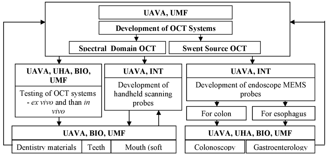 The training of these PGs is therefore a challenge [5], especially since they have to move occasionally from one institution to another, and there are various aspects they have to approach by working in small groups at a time. These aspects are covered normally by the coordinator of the project at each institution, but even that has in this case not proved sufficient, and therefore we have designated actually a deputy for each coordinator to be able to face the very diverse tasks that appear. For such a multidisciplinary work, one has to learn not only the scientific component, but also components related to the management of the activities – and this happens at all levels. Although the project has started for less than a year, most of the PG and UG students involved have participated in a conference related to the topics approached – and this has been a specific part of our strategy for their development. For some of them (maily for the UGs) attending this conference has been a very early research experience, while for some of them (even for some PGs) it was the first time that they participated in an international conference (the LASER Congress in Munchen was such a conference). This prepares the ground for more complete journal papers to conclude their various directions of work – from optomechatronics to automation, medicine (endoscopy, gastroenterology) and dentistry (with various aspects, from materials studies in prosthetics to the imaging of the soft tissue of the mouth). It provides the students with a feedback on the level of their work, while also giving them the right image on what the (expected) level of the reseach is internationally. The three years time interval of the project are planned to allow for the complete development of each research theme, but we have verified in practice that a good start is a must for success. Another most interesting aspect of PG training in our Consortium – particularly, but not only on this project - is the fact that the students (but the seniors as well) come into contact with aspects well outside their usual area of expertise. Thus, everyone has to learn and to expand his/her view to an inter-disciplinarity that may prove quite useful in their future or current carreer. 4.R&D, TRAINING AND TEACHING RELATED TO RESEARCH AT EACH PARTNER
For our current project we approach GS-based handheld probes [45], as well as endoscopic miniature scanning heads. Another research avenue is on the optimal scanning algorithms that have to be employed in OCT (but more general, in Biomedical Imaging) to obtain the most – in terms of speed (for in vivo real-time imaging) and artifact-free images – from the OCT systems. The translation of the research-gained expertise in UG teaching has been presented in detail in [5], while a most extended discussion on the methodological aspects has been done in [46].
The AOG has pioneered en face Optical Coherence Tomography (OCT) technology in 1996 and has produced the first OCT en face images from the retina, has researched and protected the generation of a combined OCT / confocal image [48], generated 3D images from retina and skin, devised multi-interferometer configurations and special modulators to collect simultaneously images from different depths in the tissue and introduced the concept of OCT imaging with adjustable depth resolution. AOG also performed theoretical studies on the performances of OCT systems. First images of eye with pathologies using en-face OCT have been reported. AOG has also developed the first instrument combining OCT with ICG fluorescence for imaging the retina in collaboration with New York Eye and Ear Infirmary and Ophthalmic Technology Inc. Combination of three technologies in one system, OCT, scanning laser ophthalmoscopy and adaptive optics and highest limit in vivo obtained in en-face images as thin as 3 μm were reported. Resources were oriented towards extending the expertise acquired from ophthalmology to cell imaging and OCT applicability to image embryos [49] and more recently, towards OCT endoscopy [45]. Expertise was extended to dentistry [47], to imaging of basal cell carcinoma and to art conservation [50]. Other current research focuses on a novel solutions to reject the effect of mirror terms in spectrometer based OCT [51], to eliminate the speckle in OCT, to perform 3D imaging using multiple paths configurations [52, 53], on coherence gated wavefront sensors [54] and on speeding up the acquisition based on graphics cards [55]. Professor Podoleanu has been involved in supervision of postdoctoral researchers constantly from 1999, with a number fluctuating, up to 5/year and of PhD students, with a number up to 8/year. He has created a new lecture course, required to be taught to students recruited by a Marie Curie Training site, HIRESOMI (2006 – 20010) he has coordinated with two other universities and three companies in Europe, supported by the EC, Biomedical Optic. This is now being taught to other PhD students in the School of Physical Sciences in the UoKent. Research inspired undergraduate projects are conducted by the AOG with students from the 3rd year MSc (PH600) and 4th year MPhys (PH700) in the School of Physical Sciences. They cover several aspects of applied optics, such as designing hand held probes for imaging the eye, for imaging in the mouth, for endoscopy and addressing fundamental limits in the technology, such as devising simple methods for linearisation of data before FFT [56]. An applied optics project is conducted with students from the 3rd year Forensic Science programs on investigating the OCT as an anti-spoof tool in imaging fingerprint at security points. Other projects are conducted with students from other universities in the South East of England, supported for short summer projects by South East Physics Network (SEPNET), embracing assembly of optical configurations for OCT as well as devising customised LabView Matlab programmes for OCT interfaces. AOG is also regularly acting as host of Erasmus work placement students, sent by Universities in Brno, Lille and Porto for 3 – 10 months to develop practical skills in an optics lab. Such exchanges resulted in published results in journals [57] and several conferences.
Since 2007, research in the ODALab started focused efforts in freeform optics driven by critical needs in compact and mobile instrumentation, specifically, in the applications of AR [75-76] and biomedical research. Several national and international sponsors, collaborators and industrial partners participate in the research efforts. Support is given through grants from the National Science Foundation, the NYSTAR Foundation, the II-VI Foundation, government research labs and industry. As part of a growing interest in freeform optics [77], Prof. Rolland is currently the director of a planned NSF Center for Freeform Optics, collaboration between UoR and the University of North Carolina Charlotte. In terms of UG students at the University of Rochester, there is a Journal of Undergraduate Research (JUR), entirely focused on publishing peer-review articles, from all domains, but only from UG students. Also, some half of the UG and most PG (Master) students are implied in research. Prof. Rolland, by example, is the Director of the Robert E. Hopkins Center for Optical Design an Engineering that was created to provide students with a computer lab, a fabrication lab, and a testing lab, that all, from UG to PG can access to develop hands on experience. Activities with the Center are integrated as part of lectures to provide a place for experiencing hands on learning. Also several classes have a lab component to give students hands-on experience with knowledge taught in the classroom. Training of PG (Doctoral students, but also PostDocs and Master students) is an essential component of the ODALab activity, and the funding discussed previously is focused to support their activity. There are thus up to 20 young researchers at a time in Prof. Rolland’s supervision – over half are Ph.D. students. The collaboration of the Consortium with ODALab has started in 2009, when Prof. Duma (UAVA) was a Fulbright Senior Research Fellow at The Institute of Optics, University of Rochester, in Sept. 2009-June 2010. It continues on issues of scanning in OCT [35], especially for galvoscanners [44], as well as on various problems of optical engineering.
The activity in the Nanofabrication Facility is focused on the design, manufacturing and testing of Micro-Electrical-Mechanical Systems (MEMS). The collaboration of the Facility with the Consortium (Fig. 1) is focused on the manufacturing and testing of MEMS for different types of endoscopes for OCT. In terms of teaching, the main interests of the Facility head, Prof. Ioana Voiculescu are to teach the students at UG and PG about emerging technologies. The emergence of new technologies that are revolutionizing the practice of engineering, the miniaturization of mechanical device, the advent of nanotechnology, the emergence of intelligent systems, the introduction of new and advanced materials, the development of sophisticated software and finally the revolution in biology are the base for modeling a modern curriculum in the Mechanical Engineering studies [79]. Based on these considerations the teaching is focused on the study of MEMS technology with a focus on optics applications. For the study of MEMS device a special commercial chip named Class on Chip (Class on a Chip, Inc., TX, USA) [80] is used. This chip is based on MEMS devices and provides innovative educational and research platforms for students at UG and PG level. This silicon chip has the dimensions 6.3 mm x 2.8 mm and contains 16 devices that move, allowing a wide range of experiments to be conducted. Several devices contain micromirrors (Fig. 4d). In the application from Fig. 4a-c the students learn the process to fabricate micromirrors; they can also analyze with the microscope the micromirror and the mechanical system used to actuate the micromirror. After the students are familiar with the concept of micromirror they are asked to find papers related to this topic and each student will have a short presentation of the paper selected by the student on micro-mirrors fabrication process and applications. Figure 4.Lab-on-a-chip [65] used for MEMS teaching at the City University of New York (CUNY), NY, USA: (a) view of the chip mounted; (b) micro-motor and gears whithin the chip; (c) comb-drivers; (d) Micromirror MEMS structure on chip for student education with microstructures for optical applications. 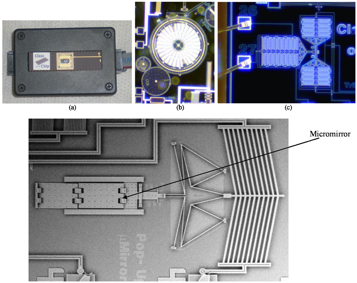 The applications in the industrial sector are increasing. There are several possible applications for MEMS and Nanotechnology in various fields like biotechnology (ex: biochips for detection of hazardous chemical and biological agents), medicine (MEMS pressure sensors) or communication (circuit) [81]. MEMS have a lot of advantages: small size and small scale allow to place a lot of different devices on the same system; it is also less expensive because we minimize the materials consumption used to make MEMS. With this kind of system you can transfer a small motion on nano-scale to a bigger motion on the macro-scale.
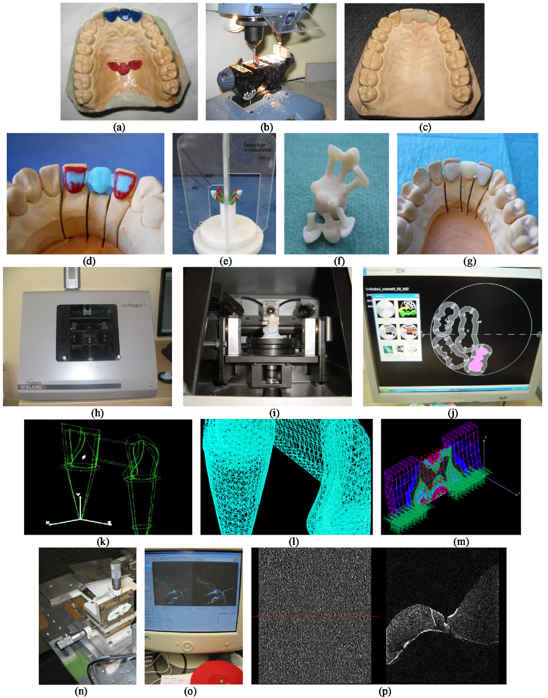
The investigation itinerary is the normal one, as biological samples are first studied in vitro, and after the calibration, in vivo. We expect that at the end of the project the OCT equipments (system & different scanning probes) will be ready to move from lab to the clinic environment. Among the research objectives targeted there are: Barrett’s esophagus and the investigation or the removal (endoscopically) of polypoid lesions. One has to stress that traditional procedures such as Ultra-magnifying Endocytoscopy, Autofluorescence Imaging, Chromoendoscopy or Confocal Laser Endomicroscopy have important advantages: they reduce unnecessary biopsies or resections, and they therefore decrease the risk of endoscopic complications. Their drawbacks (high costs, labor intensive, time consuming, and need experienced MDs) imposes OCT as a valuable tool for investigations. OCT will be used to by example to differentiate between neoplastic and non-neoplastic lesions, and to increase the accuracy of the differential diagnosis, as in colon adenoma.
5.CONCLUSIONSThe paper presents our Consortium, our main collaborators [1, 2], our current Partnership Project [4] and some of its challenges. A few aspects related to the necessary interactions between universities and industry in our Consortium (and to the role of the later) were also pointed out. These efforts are part of a longer term strategy that aims to create the structure of a future Photonics pole in Western Romania. Issues and activities related to the training of postgraduate (PG) students and to the teaching of undergraduate (UG) students (and to the translation of the research experience for the teaching process) are pointed out – at the academic partners. This paper completes – in terms of a collaborative structure – the presentation made on these aspects in [6] for the activity of the 3OM Optomechatronics Group [3]. Future work comprises further translation of the research results and expertise into the UG and Master curricula, the enhancement of our early research experinece with UGs, and a better training of the PG, especially at Doctoral and PostDoc level, with a focus on driving them towards higher impact research - and publications - at an as-early-as-possible stage of their work. Directions of development of the Photonics Pole structure are also envisaged, with the inclusion of other partners from the region and with the enhancement of the collaborations with our partners from Europe and USA. There is also a continuous, natural effort to attract significant funding for the development of our activities in R&D and to its applications in industry and medicine. ACKNOWLEDGMENTSThis work was supported by a Partnership grant of the Romanian National Authority for Scientific Research, CNDI-UEFISCDI project number PN-II-PT-PCCA-2011-3.2-1682. A. Gh. Podoleanu acknowledges the ERC 7th Framework Programme, Advanced Grant ‘COGATIMABIO’ 249889 and the NIHR Biomedical Research Centre at Moorfields Eye Hospital NHS Foundation Trust and UCL Institute of Ophthalmology. REFERENCESDuma, V. F., Negrutiu, M. L., Sinescu, C., Voiculescu, I., Miutescu, E., Burlea, A., Vlascici, M., Podoleanu, A. Gh.,
“Research lead student projects on multi-disciplinary optomechatronics with applications in biomedical imaging,”
New Trends in Educational Activity in the Field of Mechanism and Machine Theory, 19 145
–153
(2013). https://doi.org/10.1007/978-3-319-01836-2 Google Scholar
Duma, V. F., Mnerie, C., Demian, D., Hutiu, G., and Kaposta, I.,
“Building an Optomechatronics Group in a young university in Western Romania,”
in Proc. SPIE,
ET100-93
(2013). Google Scholar
Marshall, G. F., Handbook of Optical and Laser Scanning, 417
–476 Marcel Dekker, New York
(2004). Google Scholar
McDermid, I. S., Beyerle, G., Haner, D. A., and Leblanc, T.,
“Redesign and improved performance of the tropospheric ozone lidar at the jet propulsion laboratory table mountain facility,”
Applied Optics, 41 7550
–7555
(2002). https://doi.org/10.1364/AO.41.007550 Google Scholar
He, Y., Jin, W., Liu, G., Gao, Z., Wang, X., and Wang, L.,
“Modulate chopper technique used in pyroelectric uncooled focal plane thermal imager,”
in Proc. SPIE,
283
–288
(2002). Google Scholar
Duma, V. F.,
“Theoretical approach on optical choppers for top-hat light beam distributions,”
J. of Optics A: Pure and Applied Optics, 10 064008
(2008). https://doi.org/10.1088/1464-4258/10/6/064008 Google Scholar
Duma, V. F.,
“Optical choppers with circular-shaped windows: Modulation functions,”
Communications in Nonlinear Science and Numerical Simulation, 16
(5), 2218
–2224
(2011). https://doi.org/10.1016/j.cnsns.2010.04.043 Google Scholar
Duma, V. F.,
“Prototypes and modulation functions of classical and novel configurations of optical chopper wheels,”
Latin American Journal of Solids and Structures, 10
(1), 5
–18
(2013). https://doi.org/10.1590/S1679-78252013000100003 Google Scholar
Cira, O. and Duma, V. F.,
“Transmission functions of optical choppers for Gaussian beam distributions: Modeling and simulations,”
in Proc. SPIE,
87890E
(2013). Google Scholar
Marshall, G. F., Handbook of Optical and Laser Scanning, CRC Press, New York
(2011). Google Scholar
Duma, V. F.,
“On-line measurements with optical scanners: metrological aspects,”
in Proc. SPIE,
606
–617
(2005). Google Scholar
Beiser, L.,
“Design equations for a polygon laser scanner,”
in Proc. SPIE,
60
–66
(1991). Google Scholar
Duma, V. F.,
“Novel approaches in the designing of polygon scanners,”
in Proc. SPIE,
6785
–1Q
(2007). Google Scholar
Aylward, R. P.,
“Advances and technologies of galvanometer-based optical scanners,”
in Proc. SPIE,
158
–164
(1999). Google Scholar
Montagu, J.,
“Scanners - galvanometric and resonant,”
Encyclopedia of Optical Engineering, 2465
–2487
(2003). Google Scholar
Duma, V. F.,
“Optimal scanning function of a galvanometer scanner for an increased duty cycle,”
Optical Engineering, 49
(10), 103001
(2010). https://doi.org/10.1117/1.3497570 Google Scholar
Duma, V. F.,
“Command functions of open loop galvanometer scanners with optimized duty cycles,”
Theoretical and Applied Mechanics Letters, 2
(4), 043005
(2012). https://doi.org/10.1063/2.1204305 Google Scholar
Duma, V. F. and Mnerie, C.,
“Optimization of scanning and command functions of galvanometer-based scanners,”
in Proc. SPIE,
8083
–45
(2011). Google Scholar
Mnerie, C., Preitl, S., and Duma, V. F.,
“Mathematical model of a galvanometer-based scanner: simulations and experiments,”
in Proc. SPIE,
8789
–43
(2013). Google Scholar
Yang, Y.,
“Analytic solution of free space optical beam steering using Risley prisms,”
J. Lightwave Technol., 26 3576
–3583
(2008). https://doi.org/10.1109/JLT.2008.917323 Google Scholar
Warger II, W. C. and DiMarzio, Ch. A.,
“Dual-wedge scanning confocal reflectance microscope,”
Opt. Letters, 32 2140
–2142
(2007). https://doi.org/10.1364/OL.32.002140 Google Scholar
Garcia-Torales, G., Strojnik, M. and Paez, G.,
“Risley prisms to control wave-front tilt and displacement in a vectorial shearing interferometer,”
Appl. Opt., 41 1380
–1384
(2002). https://doi.org/10.1364/AO.41.001380 Google Scholar
Schitea, A., Tuef, M., and Duma, V. F.,
“Modeling of Risley prisms devices for exact scan patterns,”
in Proc. SPIE,
8789
–40
(2013). Google Scholar
Duma, V. F.,
“Radiometric versus geometric, linear and non-linear vignetting coefficient,”
Applied Optics, 48
(32), 6355
–6364
(2009). https://doi.org/10.1364/AO.48.006355 Google Scholar
Duma, V. F. and Nicolov, M.,
“Neutral density filters with Risley prisms: analysis and design,”
Appl. Opt., 48 2678
–2685
(2009). https://doi.org/10.1364/AO.48.002678 Google Scholar
Richter, B.,
“Laser scan devices for industrial application,”
WIRE, 42
(6), 529
–540
(1992). Google Scholar
Duma, V. F.,
“Dimensional measurements with optical scanners,”
in Proc. 4th European Workshop on Structural Health Monitoring (ESHM),
1217
–1224
(2008). Google Scholar
Huang, D., Swanson, E. A., Lin, C. P., Schuman, J. S., Stinson, W. G., Chang, W., Hee, M. R., Flotte, T., Gregory, K., Puliafito, C. A., and Fujimoto, J. G.,
“Optical coherence tomography,”
Science, 254
(5035), 1178
–1181
(1991). https://doi.org/10.1126/science.1957169 Google Scholar
Wojtkowski, M.,
“High-speed optical coherence tomography: basics and applications,”
Applied Optics, 49 D30
–D61
(2010). https://doi.org/10.1364/AO.49.000D30 Google Scholar
Duma, V.F.,
“Optomechatronic Modulators: Choppers, Attenuators and Scanners. Analysis and Design,”
(2013). Google Scholar
Duma, V. F., Rolland J. P., and Podoleanu, A. Gh.,
“Perspectives of optical scanning in OCT,”
in Proc. SPIE,
7556
–10
(2010). Google Scholar
Yun, S. H., Boudoux, C., Tearney, G. J., and Bouma, B. E.,
“High-speed wavelength-swept semiconductor laser with a polygon-scanner-based wavelength filter,”
Optics Letters, 28 1981
–1983
(2003). https://doi.org/10.1364/OL.28.001981 Google Scholar
Oh, W. Y., Yun, S. H., Tearney, G. J., and Bouma, B. E.,
“115 kHz tuning repetition rate ultrahigh-speed wavelength-swept semiconductor laser,”
Opt. Letters, 30 3159
–3161
(2005). https://doi.org/10.1364/OL.30.003159 Google Scholar
Mao, Y., Flueraru, C., Sherif, S., and Chang, S.,
“High performance wavelength-swept laser with mode-locking technique for optical coherence tomography,”
Optics Communications, 282 88
–92
(2009). https://doi.org/10.1016/j.optcom.2008.09.059 Google Scholar
Duma, V. F. and Podoleanu, A. Gh.,
“Theoretical approach on a galvanometric scanner with an enhanced duty cycle,”
in Proc. SPIE,
71390D
(2008). Google Scholar
Li, Y.,
“Beam deflection and scanning by two-mirror and two-axis systems of different architectures: a unified approach,”
Appl. Opt., 47 5976
–5985
(2008). https://doi.org/10.1364/AO.47.005976 Google Scholar
Duma, V. F.,
“Mathematical Functions of a 2-D Scanner with Oscillating Elements,”
Modeling, Simulation and Control of Nonlinear Engineering Dynamical Systems, 243
–253
(2009). https://doi.org/10.1007/978-1-4020-8778-3 Google Scholar
Kim, K. H., Buehler, C., and So, P. T. C.,
“High-speed, two-photon scanning microscope,”
Appl. Opt., 38 6004
–6009
(1999). https://doi.org/10.1364/AO.38.006004 Google Scholar
Duma, V. F. and Podoleanu, A. Gh.,
“Polygon mirror scanners in biomedical imaging: a review,”
in Proc. SPIE,
8621V
(2013). Google Scholar
Duma, V. F., Lee K.-S., Meemon P., and Rolland, J. P.,
“Experimental investigations of the scanning functions of galvanometer-based scanners with applications in OCT,”
Applied Optics, 50
(29), 5735
–5749
(2011). https://doi.org/10.1364/AO.50.005735 Google Scholar
Cernat, R., Tatla, T. S., Pang, J., Tadrous, P. J., Bradu, A., Dobre, G., Gelikonov, G., Gelikonov, V., and Podoleanu, A. Gh.,
“Dual instrument for in vivo and ex vivo OCT imaging in an ENT department,”
Biomed. Opt. Express, 3 3346
–3356
(2012). https://doi.org/10.1364/BOE.3.003346 Google Scholar
Duma, V. F.,
“Teaching Mechanisms: from Classical to Hands-on-Experiments and Research-Oriented,”
Mechanisms and Machine Science, 5
(8), 493
–501
(2010). Google Scholar
Sinescu, C., Negrutiu, M. L., Todea, C., Balabuc, C., Filip, L., Rominu, R., Bradu, A., Hughes, M. and Podoleanu, A. Gh.,
“Quality assessment of dental treatments using en-face optical coherence tomography,”
J. Biomed. Opt., 13
(05), 054065
(2008). https://doi.org/10.1117/1.2992593 Google Scholar
Podoleanu, A. Gh. and Rosen, R. B.,
“Combinations of techniques in imaging the retina with high resolution,”
Progress in Retinal and Eye Research, 27 464
–499
(2008). https://doi.org/10.1016/j.preteyeres.2008.03.002 Google Scholar
Ma, L., Bradu, A., Podoleanu, A. Gh., and Bloor, J. W.,
“Arrhythmia Caused by a Drosophila Tropomyosin Mutation Is Revealed Using a Novel Optical Coherence Tomography Instrument,”
PLoS, 5
(12), 1
–8
(2010). Google Scholar
Liang, H., Cid, M. G., Cucu, R. G., Dobre, G. M., Podoleanu, A. Gh., Pedro, J., and Saunders, D.,
“En-face optical coherence tomography - a novel application of non-invasive imaging to art conservation,”
Opt. Express, 13
(16), 6133
–6144
(2005). https://doi.org/10.1364/OPEX.13.006133 Google Scholar
Podoleanu, A. Gh.,
“Unique interpretation of Talbot bands and Fourier domain white light interferometry,”
Opt. Express, 15
(15), 9867
–9876
(2007). https://doi.org/10.1364/OE.15.009867 Google Scholar
Rogers, J. A., Bradu, A., and Podoleanu, A. Gh.,
“Polarization maintaining multiple-depth en face optical coherence tomography system using active re-circulation loops in the non-stationary state,”
Opt. Express, 20 29196
–29209
(2012). https://doi.org/10.1364/OE.20.029196 Google Scholar
Zurauskas, M., Rogers, J., and Podoleanu, A. Gh.,
“Simultaneous multiple-depths en-face optical coherence tomography using multiple signal excitation of acousto-optic deflectors,”
Opt. Express, 21 1925
–1936
(2013). https://doi.org/10.1364/OE.21.001925 Google Scholar
Wang, J. and Podoleanu, A. Gh.,
“Demonstration of real-time depth-resolved Shack-Hartmann measurements,”
Opt. Letters, 37
(23), 4862
–4864
(2012). https://doi.org/10.1364/OL.37.004862 Google Scholar
Van der Jeught, S., Bradu, A., and Podoleanu, A. Gh.,
“Real-time resampling in Fourier domain OCT using a graphics processing unit,”
JBO Letters, 15 030511
(2010). Google Scholar
Payne, A. and Podoleanu, A Gh.,
“Direct electronic linearization for camera-based spectral domain optical coherence tomography,”
Opt. Letters, 37
(12), 2424
–2426
(2012). https://doi.org/10.1364/OL.37.002424 Google Scholar
Bouchal, P., Bradu, A., and Podoleanu, A. Gh.,
“Gabor fusion technique in a Talbot bands optical coherence tomography system,”
Opt. Express, 20
(5), 5368
–5383
(2012). https://doi.org/10.1364/OE.20.005368 Google Scholar
Robinett, W. and Rolland, J. P.,
“A computational model for the stereoscopic optics of a head-mounted display,”
Presence: Teleoperators and Virtual Environments (MIT Press), 1
(1), 45
–62
(1992). https://doi.org/10.1162/pres.1992.1.1.45 Google Scholar
Rolland, J. P.,
“Wide angle, off-axis, see-through head-mounted display,”
Optical Engineering, 39
(7), 1760
–1767
(2000). https://doi.org/10.1117/1.602555 Google Scholar
Cakmakci, O. and Rolland, J. P.,
“Head-worn displays: A review,”
IEEE/OSA Journal of Display Technology, 2
(3), 199
–216
(2006). https://doi.org/10.1109/JDT.2006.879846 Google Scholar
Rolland, J. P., Ariely, D., and Gibson, W.,
“Towards quantifying depth and size perception in virtual environments,”
Presence: Teleoperators and Virtual Environments, 4
(1), 24
–49
(1995). https://doi.org/10.1162/pres.1995.4.1.24 Google Scholar
Rolland, J. P., Meyer, C., Arthur, K., and Rinalducci, E.,
“Methods of adjustments versus method of constant stimuli in the quantification of accuracy and precision of rendered depth in head-mounted displays,”
Presence: Teleoperators and Virtual Environments, 11
(6), 610
–625
(2002). https://doi.org/10.1162/105474602321050730 Google Scholar
Biocca, F. and Rolland, J. P.,
“Virtual eyes can rearrange your body: adaptation to virtual eye location in see-thru head-mounted displays,”
Presence: Teleoperators and Virtual Environments, 7
(3), 262
–277
(1998). https://doi.org/10.1162/105474698565703 Google Scholar
Lee, K.-S. and Rolland, J. P.,
“Bessel-Beam Spectral-Domain High-Resolution OCT with Microoptics Axicon Providing Extended Focusing Range,”
Optics Letters, 33
(15), 1696
–1698
(2008). https://doi.org/10.1364/OL.33.001696 Google Scholar
Murali, S., Lee, K.-S., and Rolland, J. P.,
“Invariant resolution dynamic focus OCM based on liquid crystal lens,”
Opt. Express, 15 15854
–15862
(2007). https://doi.org/10.1364/OE.15.015854 Google Scholar
Murali, S., Thompson, K. P., and Rolland, J. P.,
“Three-dimensional adaptive microscopy using embedded liquid lens,”
Opt. Letters, 34 145
–147
(2009). https://doi.org/10.1364/OL.34.000145 Google Scholar
Murali, S., Panomsak, M., Lee, K.-S., Kuhn, W. P., Thompson, K. P., and Rolland, J. P.,
“Assessment of a liquid lens enabled in vivo optical coherence microscope,”
Applied Optics, 49
(16), D145
–156
(2010). https://doi.org/10.1364/AO.49.00D145 Google Scholar
Rolland, J. P., Meemon, P., Murali, S., Thompson, K. P., and Lee, K.-S.,
“Gabor-based fusion technique for Optical Coherence Microscopy,”
Opt. Express, 18 3632
–3642
(2010). https://doi.org/10.1364/OE.18.003632 Google Scholar
Lee, K.-S., Thompson, K. P., and Rolland, J. P.,
“Broadband astigmatism-corrected Czerny-Turner spectrometer,”
Optics Express, 18
(22), 23378
–23384
(2010). https://doi.org/10.1364/OE.18.023378 Google Scholar
Lee, K.-S., Thompson, K. P., Meemon, P., and Rolland, J. P.,
“Cellular resolution optical coherence microscopy with high acquisition speed for in-vivo human skin volumetric imaging,”
Opt. Letters, 36 2221
–2223
(2011). https://doi.org/10.1364/OL.36.002221 Google Scholar
Lee, K.-S, Zhao, H., Ibrahim, S. F., Meemon, N., Khoudeir, L., and Rolland, J. P.,
“Three-dimensional imaging of normal skin and nonmelanoma skin cancer with cellular resolution using Gabor domain optical coherence microscopy,”
J. Biomed. Opt., 17
(12), 126006
(2012). https://doi.org/10.1117/1.JBO.17.12.126006 Google Scholar
Meemon, P., Lee, K.-S., and Rolland, J. P.,
“Doppler imaging with dual-detection full-range frequency domain optical coherence tomography,”
Biomed. Opt. Express, 1
(2), 537
–552
(2010). https://doi.org/10.1364/BOE.1.000537 Google Scholar
Meemon, P. and Rolland, J. P.,
“Swept-source based, single-shot, multi-detectable velocity range Doppler optical coherence tomography,”
Biomed. Opt. Express, 1
(3), 955
–966
(2010). https://doi.org/10.1364/BOE.1.000955 Google Scholar
Cakmakci, O. and Rolland, J. P.,
“Design and Fabrication of a Dual-Element Off-Axis Near-Eye Optical Magnifier,”
Optics Letters, 11
(32), 1363
–1365
(2007). https://doi.org/10.1364/OL.32.001363 Google Scholar
Cakmakci, O., Moore, B., Foroosh, H., and Rolland, J. P.,
“Optical local shape description for rotationally non-symmetric optical surface design and analysis,”
Optics Express, 16
(3), 1583
–1589
(2008). https://doi.org/10.1364/OE.16.001583 Google Scholar
Thompson, K. P. and Rolland, J. P.,
“Freeform Optical Surfaces – A Revolution in Imaging Optical Design,”
Optics and Photonics News, 30
–37
(2012). https://doi.org/10.1364/OPN.23.6.000030 Google Scholar
Duma, V. F. and Rolland, J. P.,
“Mechanical Constraints and Design Considerations for Polygon Scanners,”
Mechanisms and Machine Science, 5
(8), 475
–483
(2010). Google Scholar
Delale, F., Liaw, B.M., Jiji, L. M., Voiculescu, I., and Yu, H.,
“Infusion of Emerging Technologies and New Teaching Methods into The Mechanical Engineering Curriculum at The City College of New York,”
Advances in Engineering Education, 2
(4),
(2011). Google Scholar
Voiculescu, I., Zaghloul, M. E., McGill, R. A., Houser, E. J., and Fedder, G. K.,
“Electrostatically actuated resonant microcantilever beam in CMOS technology for the detection of chemical weapons,”
IEEE Sensors Journal, 5
(4), 641
–647
(2005). https://doi.org/10.1109/JSEN.2005.851016 Google Scholar
|