|
|
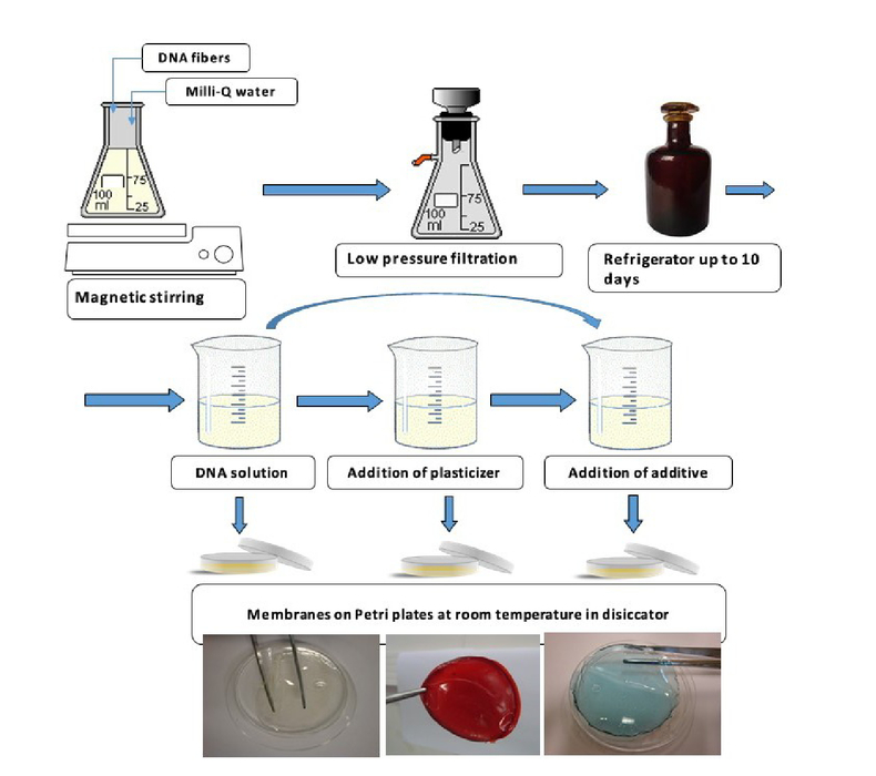
1.INTRODUCTIONBio-inspired materials are obtained from natural molecules or macromolecules. They are environmentally friendly and can be as good as synthetic ones when applied in electrochemical devices. The main advantage of natural macromolecules is that they are obtained not only from fast growing plants, but also from agricultural or fish industry waste, ex., sugar cane bagasse, shrimp carapace, fruit skins, etc. The other advantage is that they are biocompatible, what is promptly used in food, pharmaceutics and cosmetic industries. Finally, they are biodegradable, what is very important to preserve our environment. The most important natural macromolecules are polysaccharides, proteins, DNA and natural rubber. Beside the cellulose-paper industry, other polysaccharides are commonly used in food and in pharmaceutic industries. Their derivatives are good additives for cosmetic products. Proteins are used principally in textiles, and natural rubber in automotive industry. For instance, DNA extracted from fish industry waste is not yet a commodity for any industry. However, as already shown in several papers it is a promising material that can be used in opto-electronic application [1-3] as wave guides [4-5], ionically conducting membranes for electrochemical devices such as electrochromic (ECD) [6], dye sensitized solar cells (DSSC), organic filed-effect transistors (OFET) [7-8] or organic light emitting diodes (OLED) [9]. The solubility of materials is very important for the electronics industry because allows to use less expensive deposition methods as for example roll-to-roll process. Natural macromolecules are mostly water soluble and so is DNA, and this can be a problem for some applications. However, it is possible to improve their physicochemical properties through chemical or physical derivatizations, i.e., by grafting, crosslinking or plasticization processes. These reactions lead to solubility, gelatinization and retrogradation (recrystallization) property changes of these macromolecules. As an example, substitution of DNA sodium counter ions, from salmon sperm extraction, by cetyl trimethyl ammonium (CTMA) [10] or dioctadecyl dimethyl ammonium (DODA) [11] cations results in alteration of DNA solubility from water to low polar organic solvents such as butanol [5]. Addition of glycerol, which is widely used as a plasticizer for natural polymers, promotes a separation of the polymeric chains through the Van der Waals forces or hydrogen bonds formation because the polymer-polymer and polymer-plasticizer interactions [12]. As mentioned above DNA is an interesting natural material that when having a high molecular weight then can easily form membranes with excellent optical properties [13-16]. DNA have the structure of heteroatoms, which could complex the lithium ions, protons or anions, or even release protons or sodium ions and thereby promote ionic conduction in these systems [17-18]. It can show ionic conduction properties in association with ionic liquids [19] or quasi reversible electrochromic properties when containing alkyl viologen [20]. Tests of application of DNA in OLEDs were also reported [21]. It was shown that using modified, soluble in organic solvents DNA, can improve the electroluminescent properties of OLEDs [21]. DNA-CTMA lipid complex was used in an OLED as an active charge transport layer aiming to elucidate the semiconducting characteristics of this material [22]. The obtained results revealed that a DNA-CTMA layer preferentially transports holes rather than electrons. Furthermore, in using sandwiched device structures with the DNA-CTMA layer, it was observed that the DNA-CTMA layer basically possess both hole and electron transport abilities. The preferential hole transport, which is due to a shallow lowest unoccupied molecular orbital (LUMO) level, prohibits an efficient electron injection from an adjacent carrier transport layer. In this communication, we present some of our results of synthesis and application of DNA-based membranes in electrochromic windows (ECDs) and dye sensitized solar cells (DSSC). 2.EXPERIMENTALThe DNA-based membranes were obtained by dispersion of DNA in water and/or Milli-Q® water/etanol 1:1 solution and further addition of additives such as lithium salts, acids, rare earth compounds such as Eu(CF3SO3)3 and Er(CF3SO3)3 (Sigma Aldrich, 98%), glycerol (Himedia, 99.5%) etc. In our studies, we used salmon sperm DNA either in its sodium salt (DNAO; molecular weight of about 8×10⁶ g/mol; Ogata Research Laboratory, Chitose Institute of Technology, Japan) or acid (DNAA; Aldrich®) form. As the DNAo had much higher molecular weight than DNAA, about 3 days were needed to dissolve it in water. After complete dissolution of DNA, some small quantities were stored in hermetically closed glass flasks at room temperature. The rest was versed on Petri plates and cast for 72 h at room temperature until the complete dryness. Rare earth compound samples were additionally dried in an oven at 60 °C for 8 to 12 days [23-24]. The samples preparation is shown in Figure 1. The samples of DNA-PEDOT:PSS and DNA-PB were obtained by addition of either PEDOT:PSS or Prussian Blue (PB) to the above described DNA solution. The PEDOT:PSS was of 5% of DNA mass. The thickness of resulting membranes were of about 5 x 10⁻³ cm. Complex impedance measurements were performed on membranes with the disc shape that were sandwiched between two stainless-steel electrodes and placed under reduced pressure in a hermetically closed Teflon holder. Impedance data were collected with a Solartron model 1260 using an AC potential of 50 mV in the temperature range of 25 to 75 °C and frequency range of 10 Hz to 1 MHz. The electrochromic devices (ECDs) with 1 to 2 cm2 area and having the glass/ITO/WO3/DNA-based electrolyte/CeO2-TiO2/ITO/glass configuration were obtained by assembling glass/ITO/WO3 and CeO2-TiO2/ITO/glass pieces. One cm free space was left for the electrical contact. The DNA-membrane was placed in such a way that the two coatings faced each other inside the assembled window. A 1 cm wide Cu-conducting tape (3 M) was glued to the free edge of each substrate for electrical connections. The mounted cells were finally sealed with a protective and insulating tape (3 M). The DSSC were assembled using transparent, conducting glass/FTO (fluor tin oxide; Hartford; glass, ≤30 Ω/cm2) substrates previously washed with water and detergent, and next cleaned in ultrasound bath for 15 min in acetone, isopropanol, ethanol and distilled water each. Then the substrates were dried with dry N2. Aiming to control the TiO2 thickness three layers of adhesive tape (Scotch Magic Tape 3M® with 50 μm of thickness) were glued parallel on both sides of glass/FTO substrate leaving 8 mm of distance between them. TiO2 colloidal solution (Ti-Nanoxide T, Solaronix) was deposited by doctor blade method on glass/FTO substrates [25]. After drying in air for few minutes the films were heat treated at 450 °C for 30 min and at heating rate of 10 °C/min in EDG3P - S owen resulting in about 30 μm thick films. The glass/FTO/TiO2-DNA or glass/FTO/TiO2-DNA-PEDOT:PSS were obtained by dipping the glass/FTO/TiO2 substrate in either DNA or DNA-PEDOT:PSS solution for 20 min. After that, these assemblies were covered with Petri plat for 2 min to promote better macromolecule penetration on TiO2-dye microporous structure. Soon after, the samples were immersed in 1.0x10⁻³ mol/L solution of hydrophobic TG6 dye [26] for 18 h. After that, some drops of polymer electrolyte based on poly(ethylene oxide-co-2-(2-methoxyethoxy) ethyl glicylether (P(EO/EM); Daiso); 70 wt% of v-butirolactone; 5 wt% of LiI and I2 [27] or DNA-CTMA or DNA-DODA solution were placed on the top. Finally, counter-electrodes (CE), prepared by sputtering a thin Pt film 400 Rs≤10 Ω/cm2) on FTO substrates, were pressed against the electrolyte coating. The DSSC devices with effective area of 0.25 cm2 (inset Fig. 3) were characterized on an optical bench consisting of an Oriel Xe (Hg) 250 W lamp with intensity of 100 mW/cm2, beam collimation lenses and filter AM 1.5. The light intensity was measured with a Newport Optical Power Meter 1830-C. Current–potential curves (I–V curves) were obtained using linear sweep voltammetry at 10 mV/s with the Eco Chimie-Autolab PGSTAT 10 potentiostat/galvanostat. The measurements were performed after 15 min of cell stabilization. The UV–Vis spectroscopy measurements in transmittance mode of DNA and DNA-PEDOT:PSS liquid samples using a 1 cm large quartz cubes were recorded with a Jasco 630V or Agilent spectrophotometer between 190 and 1100 nm. 3.RESULTS AND DISCUSSIONElectrochemical devices (ECDs) and dye-sensitized solar cells (DSSCs) require electrolytes for their functioning. Common electrolytes are liquid, but for the device application, they are not very convenient in manufacturing process. The other inconvenience is that they can leak during the device damage. Thus, since 1970’s researchers are trying to substitute the liquid electrolytes by gel or solid ones principally based on poly(ethylene oxide) (PEO) [28]. Later, other macromolecules including natural ones having heteroatoms in their structure started to be also investigated [29]. More recently, DNA-based membranes were prepared and analyzed by electrochemical and spectroscopic analysis [30]. Figure 2 shows the results of the ionic conductivity as a function of temperature of the membranes of DNA, DNA with PEDOT and modified DNA with either CTMA or DODA. As one can observe, similarly to other ionically conducting samples [31] an increase of conductivity with temperature is observed for all membranes. From this figure, it is also seen that three samples, i.e., DNA, DNA-DODA and DNA-PEDOT:PSS have almost the same ionic conductivity values at room temperature, of about 2×10⁻⁵ S/cm. The sample of DNA-CTMA-LiI/I2 shows one order of magnitude higher ionic conductivity of 5.4×10⁻⁴ S/cm. Moreover, the samples of DNA and DNA-PEDOT reveal a sharp increase of conductivity in 40 to 55 °C temperature range, which can be result of DNA denaturation process [18, 32]. From this figure it is also seen that an addition of PEDOT:PSS to the DNA promotes a decrease of the ionic conductivity of the samples, differently to the DNA-PEDOT samples with plasticizer [32]. Analyzing the ionic conductivity as a function of inverse of temperature it can be stated that all these samples display linear behavior. From the Arrhenius Equation 1, the single activation energies (Ea) of 32.2 and 37.1 kJ/mol are obtained for DNA-CTMA and DNA-DODA with 10 wt% of LiI/I2 samples, respectively. In contrary, DNA and DNA-PEDOT exhibit two Ea values. Consequently, it seems that the ionic hopping mechanism between complexation sites is predominant in these systems. Figure 2.Log of ionic conductivity as function of inverse temperature for the following membranes: DNAO (▄), DNA-CTMA-10% LiI/I2 ( 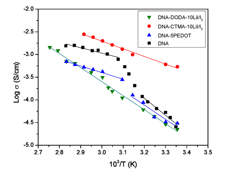 where σ0 is a pre-exponential factor proportional to the number of charge carries; Ea is the activation energy for conduction (eV); k = 8.6173 ×10⁻⁵ eV/K is the Boltzmann constant; T is the absolute temperature (K) and T is temperature in K. The DNA samples, doped with Prussian Blue (PB), Nile Blue (NB), Disperse Red 1 (DR1) or Disperse Orange (DO3) dyes display different behavior of ionic conductivity versus inverse temperature as shown in Figure 3. Except DNA-PB complex, which shows a high σ = 10-4 S/cm conductivity, all other samples exhibit significantly lower values of 10-7 S/cm. This result is most likely due to the large size of doping molecules. Prussian Blue is an inorganic compound with electrochromic properties. Thus, it changes its transmittance properties under redox conditions [35]. Moreover, these sample reveal non-linear behavior as function of inverse temperature, what shows the presence of macromolecular free volume influence on the charge carries movement [36]. Such property is frequently described by Vogel-Tamman-Fulcher (VTF) model as given in Equation 2 [37-38]. Figure 3.Log of ionic conductivity as function of inverse temperature for DNA-PB (▄), DNA-NB ( 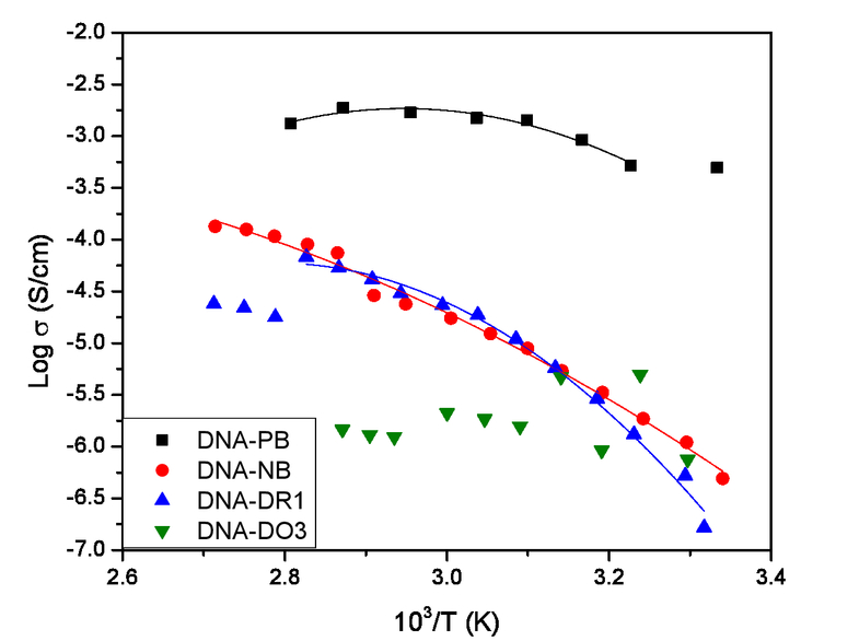 where σ0 is a pre-exponential factor proportional to the number of charge carries; Ea is pseudo-activation energy for conduction (eV); k = 8.6173×10⁻⁵ eV/K is the Boltzmann constant; T is the absolute temperature (K) and To represents ideal vitreous transition temperature, suggested to be 50 K below the glass transition temperature (Tg). Figure 4 shows the ionic conductivities for the DNA based membranes, doped with erbium and europium salts [24, 39]. From this figure one can see that the samples exhibit the ionic conductivities similar to those of PEO-doped with rare earth compounds [23]. Addition of europium salt promotes in all DNA-based samples an increase of ionic conductivity from 10-7 up to 10-4 S/cm. The DNA sample with 10% of erbium triflate (DNA-10Er) exhibits an ionic conductivity of 1.2×10⁻⁵ at 30°C and 7.8×10⁻⁴ S/cm at 100°C. That doped with 50% of europium triflate (DNA-50Eu) revealed the highest ionic conductivity of 1.0×10⁻⁴ S/cm [39]. Figure 4.Log of ionic conductivity as a function of inverse temperature of membranes of DNA-matrix (▄), DNA-50Eu (  The DNA-based membranes, doped with rare earth salts, similarly to the DNA-CTMA samples (Figure 2) evidence a linear increase of the ionic conductivities with the inverse of temperature, what indicates an Arrhenius behavior (Equation 1). The energy of activation extracted from these data for DNA-matrix, DNA-10Er and DNA-50Eu samples are of 36.14, 24.56 and 16.32 kJ/mol, respectively. It confirms that addition of either erbium or europium triflate promotes a decrease of energy of activation, probably due to the enhancement of segmental motion of the polymer chains and increase of ionic mobility. From the data shown in Figures 2-4 one can conclude that some of the DNA-based membranes fulfill the requirements of higher than 10-5 S/cm ionic conductivity, as established for polymer electrolytes to be applied in electrochemical devices [40]. Additionally, as it is seen from Figure 1, the studied here DNA-based samples can be obtained in form of transparent or colored membranes that are flexible and can stick easily to the glass or metal, what is advantageous for their application in different flexible devices. Aiming to test these membranes in electrochemical devices small ECDs were assembled, and subjected to the electrochemical and spectroscopic measurements. Figure 5 shows two examples of ECDs with glass/ITO/WO3/DNA-LiClO4/CeO2-TiO2/ITO/glass and glass/ITO/WO3/DNA-PB/CeO2-TiO2/ITO/glass configurations. The images from left to right show the ECDs in discolored, colored and discolored states, achieved by applying reversible potentials. Although the ECD with DNA-LiClO4 electrolyte exhibits a clear difference between transparent and colored states (Figure 5a) the ECD with DNA-PB seems to be always blue (Figure 5b). This permanent blue color is due to the presence of Prussian Blue dye which is intensified upon cathodic charge application and WO3 coloring. Figure 5.Images of glass/ITO/WO3/electrolyte/CeO2-TiO2/ITO/glass with DNA-LiClO4 (a) and DNA-PB (b) electrolytes.  Figure 6 displays the voltammograms of ECDs with DNA-based electrolytes. These voltammograms were performed at a scan rate of 50 mV/s with the applied potentials between -2.5 and 2.0 V for ECDs with DNA, DNA-PB and DNA-PEDOT, and in the range of -3.0 to 2.5 V for the ECD with DNA-CTMA-PB electrolyte. The obtained voltammograms show very clear cathodic and anodic peaks situated at about -1.3 and -0.5 V. As it can be seen in this figure, the voltammograms of these samples are for 200 cycles that showed the current values of -0.12 and 0.12 mA/cm2 for cathodic and anodic peaks, respectively. Similar results were obtained for windows with WO3/gelatin-LiI-I2/CeO2-TiO2 configuration [41]. The ECDs with DNA-based electrolytes show very similar voltammetries with well defined cathodic and anodic peaks. However, at this point it should be stated that depending on the electrolyte used the cyclic voltammograms change as in the case of ECDs with glass/FTO/Nb2O5:Mo/gelatin-LiClO4/CeO2-TiO2/FTO/glass [42], glass/FTO/WO3/gelatin-acetic acid/CeO2-TiO2/FTO/glass [43] or glass/FTO/WO3/gelatin-Zn(CF3SO3)2/CeO2-TiO2/FTO/glass configuration. These differences may be due to the composition of the devices, which differ in the working electrode, and the composition of electrolytes. Figure 6.Cyclic voltamograms of ECDs with electrolyte of DNA (continuous black line), DNA-PB (dashed red line), DNA-PEDOT:PSS (dotted blue line) and DNA-CTMA-PB (dash dot green line) [41].  Figure 7 shows the transmittance curves for the colored and discolored states of three ECDs with DNA-based electrolytes. As can be it an seen from this figure the transmission difference between colored and bleached states is about 8% at 550 nm, i.e., changing from 76% for transparent to 68% for colored ECD with DNA-CTMA-PEDOT electrolyte [41]. Other two devices show almost the same transmittance difference between colored and bleached states. However, the ECD with DNA-PEDOT electrolyte is darker than the others. The DNA-PEDOT electrolyte is blue colored due to the PEDOT alone, which is similar to DNA-NB membranes. As consequence, the presence of some dyes in DNA leads to already colored membranes. However, the ECD with WO3/DNA-NB/CeO2-TiO2 configuration is different of other ECDs because it starts to display a transmittance difference at about 700 nm, which in the range of 780 to 1100 nm is about 16% between color and bleached states [44]. Finally, comparing the transmittance results of ECDs with DNA-CTMA-PEDOT and DNA-PEDOT electrolytes one can observe a slightly larger difference in transmittance between the colored and the bleached states in the case of ECD with DNA-PEDOT membrane. Figure 7.UV-Vis-NIR spectra of ECDs with DNA (dashed black lines), DNA-CTMA-PEDOT:PSS (continuous blue line) and DNA-PEDOT:PSS (dotted red lines) [41].  The DNA-based membranes and solutions were used to assemble small dye sensitized solar cells (DSSC) with glass/TiO2/dye/electrolyte/Pt configuration. These DSSCs were then characterized by current-voltage dependence (I-V) measurements (Figure 8), which is the most common and powerful method to characterize DSSC [45]. From the obtained I-V curves, it is possible to obtain open circuit voltage (VOC) and short circuit current (ISC). From their shapes, one can deduce the conversion efficiency at any solar illuminations. The DNA and DNA-PEDOT were used as hole-transport-material (HTM) on glass/TiO2 substrates and the DNA-CTMA and DNA-DODA doped with LiI/I2 as electrolytes. The conversion efficiencies were obtained using the following equation: where Pmax is a maximum power, Pinc is power of incident light, which was 100 mW/cm2. In the study of DNA as HTM two kinds of DNA were used: the first one was DNA with high molecular weight (about 8 MDa), called DNAO, and the second one was DNA with low molecular weight, called DNAA. Figure 8 shows that DNAA exhibits the highest photocurrent of 2.8 mA/cm2. Addition of PEDOT to this sample promoted a decrease of photocurrent, which drop down to 2.1 mA/cm2. It is clear that the size of DNA macromolecular chains influence the DSSC performance. DNAO doped with PEDOT shows the lowest photocurrent value of 0.6 mA/cm2. The measured conversion efficiency (η) values were of 0.07% for DSSC with DNAO and 0.56% for DSSC with DNAA. The addition of 5% of PEDOT:PSS to either DNAA or DNAO formulation did not improve the conversion efficiency that dropped to 0.37 and increased to 0.08%, respectively. Figure 8.I versus V curves for DSSC with DNA-DODA-10%LiI/I2 (continuous black line), DNA-CTMA-10%LiI/I2 (dashed red line), DNAA (dash dot green line), DNAA-5% PEDOT:PSS (dotted blue line) and DNAO-5% PEDOT:PSS (dash dot dot orange line) [33-34, 46].  The fill factor of 34.11% for DNAA sample was calculated using the Equation (4). where Pmax is the maximum power, Pth is a theoretical power; Vm is a maximum potential, Jm is a maximum current JSC is a short circuit current and VOC is an open circuit voltage. As already mentioned, the best values of solar efficiency were obtained with DNAA. However, these values are comparable to DSSC with poly(epichlorohydrin-co-ethylene oxide)-based electrolyte [47] and lower when compared with other DSSC [48]. Beside this, it was observed that the DNA used in this research has a significant impact on the results. It seems that the low molecular mass DNAA purchased from Aldrich does not obstruct the charge transfer between electrolyte and TiO2 as much as high molecular mass DNAO purchased from Ogata Laboratory (Japan). 4.CONCLUSIONSThe present work describes the results of preparation and characterization of DNA-based membranes and their application in electrochemical devices. The samples of DNA and modified DNA were doped with Prussian Blue (PB), poly(ethylene dioxythiophene) (PEDOT), europium and erbium triflate and organic dyes such as Nile Blue (NB), Disperse Red 1 (DR1) and Disperse Orange 3 (DO3). The colored or colorless DNA-based membranes were then characterized by electrochemical and spectroscopic measurements and applied in electrochemical devices such as electrochromic (ECDs) or dye sensitized solar cells (DSSC). It was shown that some of the DNA-based membranes possess ionic conductivity of more than 10-5 S/cm, which is suggested for polymer electrolytes practical applications. The best results of 10-4 S/cm of ionic conductivity were obtained for DNA-CTMA, DNA-PB and DNA-Eu membranes. DNA, DNA-CTMA, DNA-DODA and DNA-PEDOT:PSS were also investigated as either hole carrier material (HTM) or polymer electrolyte in dye-sensitized solar cells (DSSC). The DNA-based samples as HTM in small DSSCs revealed a solar efficiency of 0.56%. Polymer DNA-DODA electrolyte with 10 wt% of LiI/I2 when applied in small DSSC exhibited a solar efficiency of 0.66%. The obtained results show that natural macromolecules-based membranes are not only environmentally friendly but also promising materials to be investigated for several electrochemical devices. However, to obtain better results more research is required. AcknowledgementsThe authors are indebted to FAPESP, CNPq (Proc. No. 201820/2014-5), CAPES, and European Community for FP7-PEOPLE-2009-IRSES Biomolec - 247544 and STATOIL for the financial support given to this research. REFERENCESOuchen, F., Gomez, E., Joyce, D., Williams, A., Kim, S., Heckman, E., Johnson, L., Yaney, P., Venkat, N., Steckl, A., Kajzar, F., Rau, I., Pawlicka, A., Prasad, P. and Grote, J.,
“Latest advances in bio materials: from deoxyribonucleic acid to nucleobases,”
in SPIE,
89831A1
(2014). Google Scholar
Steckl, A.J.,
“DNA - a new material for photonics?,”
Nature Photonics, 1
(1), 3
(2007). https://doi.org/10.1038/nphoton.2006.56 Google Scholar
Singh, T.B., Sariciftci, N.S. and Grote, J.G.,
“Bio-organic optoelectronic devices using DNA,”
Org. Electron, 223 189
(2010). Google Scholar
Rau, I., Grote, J., Kajzar, F. and Pawlicka, A.,
“DNA - novel nanomaterial for applications in photonics and in electronics,”
Comptes Rendus Phys., 13
(8), 853
(2012). https://doi.org/10.1016/j.crhy.2012.09.005 Google Scholar
[Optical Materials in Defence Systems Technology III], 6401 U25 2006). Google Scholar
Pawlicka, A., Grote, J., Kajzar, F., Rau, I. and Silva, M.M.,
“Agar and DNA bio-membranes for electrochromic devices applications,”
Nonlin. Optics Quant. Optics, 45 113
(2012). Google Scholar
Stadler, P., Oppelt, K., Singh, T.B., Grote, J.G., Schwodiauer, R., Bauer, S., Piglmayer-Brezina, H., Bauerle, D. and Sariciftci, N.S.,
“Organic field-effect transistors and memory elements using deoxyribonucleic acid (DNA) gate dielectric,”
Org. Electron., 8
(6), 648
(2007). https://doi.org/10.1016/j.orgel.2007.05.003 Google Scholar
Yumusak, C, Singh, T.B., Sariciftci, N.S. and Grote, J.G.,
“Bio-organic field effect transistors based on crosslinked deoxyribonucleic acid (DNA) gate dielectric,”
Appi. Phys. Lett., 95
(26), 263304
(2009). https://doi.org/10.1063/1.3278592 Google Scholar
Bazan, G.C., Zalar, P., Kamkar, D., Naik, R., Ouchen, F., Grote, J.G. and Nguyen, T.Q.,
“DNA electron injection interlayers for polymer light-emitting diodes,”
J. Am. Chem. Soc., 133
(29), 11010
(2011). https://doi.org/10.1021/ja201868d Google Scholar
Samoc, M, Samoc, A. and Grote, J.G.,
“Complex nonlinear refractive index of DNA,”
Chem. Phys. Lett., 431
(1-3), 132
(2006). https://doi.org/10.1016/j.cplett.2006.09.057 Google Scholar
Sergeyev, V.G., Pyshkina, O.A., Lezov, A.V., Mel’nikov, A.B., Ryumtsev, E.I., Zezin, A.B. and Kabanov, V.A.,
“DNA complexed with oppositely charged amphiphile in low-polar organic solvents,”
Langmuir, 15 4434
(1999). https://doi.org/10.1021/la981583h Google Scholar
Galliard, T., [Starch: Properties and Potential], John Wiley & Sons, New York
(1987). Google Scholar
Wang, L.L., Yoshida, J. and Ogata, N.,
“Self-assembled supramolecular films derived from marine deoxyribonucleic acid (DNA)-cationic surfactant complexes: Large-scale preparation and optical and thermal properties,”
Chem. Mater., 13
(4), 1273
(2001). https://doi.org/10.1021/cm000869g Google Scholar
Misoska, V., Price, W.E., Ralph, S.F., Wallace, G.G. and Ogata, N.,
“Synthesis, characterisation and ion transport studies on polypyrrole/deoxyribonucleic acid conducting polymer membranes,”
Synth. Met., 123
(2), 279
(2001). https://doi.org/10.1016/S0379-6779(01)00313-7 Google Scholar
Kawabe, Y., Wang, L., Horinouchi, S. and Ogata, N.,
“Amplified spontaneous emission from fluorescent-dye-doped DNA-surfactant complex films,”
Adv. Mater., 12
(17), 1281
(2000). https://doi.org/10.1002/(ISSN)1521-4095 Google Scholar
Grote, J.G., Hagen, J.A., Zetts, J.S., Nelson, R.L., Diggs, D.E., Stone, M.O., Yaney, P.P., Heckman, E., Zhang, C, Steier, W.H., Jen, A.K.Y., Dalton, L.R., Ogata, N., Curley, M.J., Clarson, S.J. and Hopkins, F.K.,
“Investigation of polymers and marine-derived DNA in optoelectronics,”
J. Phys. Chem. B, 108
(25), 8584
(2004). https://doi.org/10.1021/jp038056d Google Scholar
Olino, H. and Takizawa, N.,
“Ion conductive and flexible DNA films,”
Chem. Lett, 29
(6), 642
(2000). https://doi.org/10.1246/cl.2000.642 Google Scholar
Firmino, A., Grote, J.G., Kajzar, F., M’Peko, J.-C. and Pawlicka, A.,
“DNA-based ionic conducting membranes,”
J. Appi. Phys., 110 033704
(2011). https://doi.org/10.1063/1.3610951 Google Scholar
Olino, H. and Nishimura, N.,
“Ion conductive characteristics of DNA film containing ionic liquids,”
J. Electrochem. Soc., 148
(4), E168
(2001). https://doi.org/10.1149/1.1357179 Google Scholar
Kakibe, T. and Olino, H.,
“Quasi-reversible electrochromic behavior of alkyl viologens dispersed mono- molecularly in double-stranded DNA chains,”
J. Mater. Chem., 19
(28), 4960
(2009). https://doi.org/10.1039/b822818c Google Scholar
Hagen, J.A., Li, W., Steckl, J. and Grote, J.G.,
“Enhanced emission efficiency in organic light-emitting diodes using deoxyribonucleic acid complex as an electron blocking layer,”
Appi. Phys. Lett., 88
(17), 171109
(2006). https://doi.org/10.1063/1.2197973 Google Scholar
Hirata, K., Oyamada, T., Imai, T., Sasabe, H., Adachi, C. and Koyama, T.,
“Electroluminescence as a probe for elucidating electrical conductivity in a deoxyribonucleic acid-cetyltrimethylammonium lipid complex layer,”
Appi. Phys. Lett., 85
(9), 1627
(2004). https://doi.org/10.1063/1.1780600 Google Scholar
Gonçalves, A.R., da Silva, C.J.R., Silva, M.M. and Smith, M.J.,
“Ionic conduction and thermal properties of polyethylene oxide)-Er (CF3S03)(3) films,”
Ionics, 1
(4), 342
(1995). https://doi.org/10.1007/BF02390217 Google Scholar
Leones, R., Fernandes, M., Sentanin, F., Cesarino, L, Lima, J.F., Bermudez, V.d.Z., Pawlicka, A., Magon, C.J., Donoso, J.P. and Silva, M.M.,
“Ionically conducting Er3+-doped DNA-based biomembranes forelectrochromic devices,”
Electrochim. Acta, 120 327
(2014). https://doi.org/10.1016/j.electacta.2013.12.006 Google Scholar
de Freitas, J.N., Longo, C, Nogueira, A.F. and De Paoli, M.A.,
“Solar module using dye-sensitized solar cells with a polymer electrolyte,”
Sol. Energ. Mat. Sol. Cells, 92
(9), 1110
(2008). https://doi.org/10.1016/j.solmat.2008.03.022 Google Scholar
Law, C, Pathirana, S.C., Li, X., Anderson, A.Y., Barnes, P.R., Listorti, A. and Ghaddar, T.H.,
“Water-based electrolytes for dye-sensitized solar cells,”
Adv. Mater., 22
(40), 4505
(2010). https://doi.org/10.1002/adma.v22:40 Google Scholar
Benedetti, J.E.,
“Preparação, caracterização e aplicação de eletrólitos polimericos gel em células solares Ti02/corante,”
Universidade de Campinas- Unicamp,
(2010). Google Scholar
Armand, M.,
“Polymers with ionic conductivity,”
Adv. Mater., 2
(6-7), 278
(1990). https://doi.org/10.1002/(ISSN)1521-4095 Google Scholar
Regiani, A.M., Tambelli, CE., Pawlicka, A., Cúrvelo, A.A.S., Gandini, A., LeNest, J.F. and Donoso, J.P.,
“DSC and solid state NMR characterization of hydroxyethylcellulose/polyether films,”
Polym. Intern., 49
(9), 960
(2000). https://doi.org/10.1002/(ISSN)1097-0126 Google Scholar
Pawlicka, A., Firmino, A., Vieira, D., Sentanin, F., Grote, J.G. and Kajzar, F.,
“Gelatin- and DNA-based ionic conducting membranes for electrochromic devices,”
in SPIE,
74870J
(2009). Google Scholar
Pawlicka, A., Vieira, D. and Sabadini, R.C,
“Gelatin-HCl biomembranes with ionic-conducting properties,”
Ionics, 19 1723
(2013). https://doi.org/10.1007/s11581-013-0935-9 Google Scholar
Pawlicka, A., Sentanin, F., Firmino, A., Grote, J.G., Kajzar, F. and Rau, I.,
“Ionically conducting DNA-based membranes for eletrochromic devices,”
Synth. Met.,
(21-22), 1612329
(2011). Google Scholar
Jimenez, D.E.Q.,
“Síntese e estudo de membranas condutoras iónicas a base de DNA-CTMA e DNA-DODA para aplicação em células solares,”
Master, Universidade de Sao Paulo,
(2013). Google Scholar
Jayme, C.C.,
“Membranas a base de DNA e DNA-PEDOT:PSS para células solares sensibilizadas por corante,”
Master, Universidade de Sao Paulo,
(2013). Google Scholar
Bueno, P.R., Giménez-Romero, D., Ferreira, F.F., Setti, C.O., Garcia-Jareno, J.J., Agrisuelas, J. and Vicente, F.,
“Electrochromic switching mechanism of iron hexacyanoferrates molecular compounds: the role of Fe2+(CN)(6) vacancies,”
J. Phys. Chem. C, 113
(22), 9916
(2009). https://doi.org/10.1021/jp901146w Google Scholar
Liew, C.-W. and Ramesh, S.,
“Electrical, structural, thermal and electrochemical properties of corn starch-based biopolymer electrolytes,”
Carbohydrate Polym., 124
(0), 222
(2015). https://doi.org/10.1016/j.carbpol.2015.02.024 Google Scholar
Kreuer, K.D.,
“Proton conductivity: Materials and applications,”
Chem. Mater., 8
(3), 610
(1996). https://doi.org/10.1021/cm950192a Google Scholar
Kreuer, K.D.,
“On the development of proton conducting materials for technological applications,”
Solid State Ionics, 97
(1-4), 1
(1997). https://doi.org/10.1016/S0167-2738(97)00082-9 Google Scholar
Leones, R., Rodrigues, L.C, Fernandes, M., Ferreira, R.A.S., Cesarino, I., Pawlicka, A., Carlos, L.D., Bermudez, V.Z. and Silva, M.M.,
“Electro-optical properties of the DNA-Eu3+ bio-membranes,”
J. Electroanal. Chem., 708 116
(2013). https://doi.org/10.1016/j.jelechem.2013.08.031 Google Scholar
Armand, M., Duclot, M.T. and Chabagno, J.M.,
“Polymeric solid electrolytes,”
Second International Meeting on Solid State Electrolytes, 6
(1978). Google Scholar
Sentanin, F.,
“Desenvolvimento de janelas eletrocrômicas,”
PhD, Universidade de São Paulo,
(2012). Google Scholar
Avellaneda, C.O., Vieira, D.F., Al-Kahlout, A., Heusing, S., Leite, E.R., Pawlicka, A. and Aegerter, M.A.,
“Ali solid-state electrochromic devices with gelatin-based electrolyte,”
Sol. Energ. Mat. Sol. Cells, 92
(2), 228
(2008). https://doi.org/10.1016/j.solmat.2007.02.025 Google Scholar
Al-Kahlout, A., Vieira, D., Avellaneda, C.O., Leite, E.R., Aegerter, M.A. and Pawlicka, A.,
“Gelatin-based protonic electrolyte for electrochromic windows,”
Ionics, 16 13
(2010). https://doi.org/10.1007/s11581-009-0367-8 Google Scholar
Mindroiu, M., Zgârian, R.G., Kajzar, F., Rãu, L, De Oliveira, H.C.L., Pawlicka, A. and Tihan, G.T.,
“DNA-based membranes for potential applications,”
Ionics, 21
(5), 1381
(2014). https://doi.org/10.1007/s11581-014-1293-y Google Scholar
Benedetti, J.E., de Paoli, M.A. and Nogueira, A.F.,
“Enhancement of photocurrent generation and open circuit voltage in dye-sensitized solar cells using Li+ trapping species in the gel electrolyte,”
Chem. Comm., (9) 1121
(2008). https://doi.org/10.1039/b717278h Google Scholar
Jayme, C.C., Kanicki, J., Kajzar, F., Nogueira, A.F. and Pawlicka, A.,
“Influence of DNA and DNA-PEDOT:PSS on dye sensitized solar cell performance,”
Mol. Cryst. Liq. Cryst.,
(2015). Google Scholar
Nogueira, V.C., Longo, C., Nogueira, A.F., Soto-Oviedo, M.A. and De Paoli, M.A.,
“Solid-state dye-sensitized solar cell: Improved performance and stability using a plasticized polymer electrolyte,”
J. Photochem. Photobiol. a-Chem., 181
(2-3), 226
(2006). https://doi.org/10.1016/j.jphotochem.2005.11.028 Google Scholar
de Freitas, J.N., Gonçalves, A.D., de Paoli, M.A., Durrant, J.R. and Nogueira, A.F.,
“The role of gel electrolyte composition in the kinetics and performance of dye-sensitized solar cells,”
Electrochim. Acta, 53
(24), 7166
(2008). https://doi.org/10.1016/j.electacta.2008.05.009 Google Scholar
|