

|
|
1.INTRODUCTIONOptical components play an important role in high power laser systems for applications such as laser fabrication, direct energy weapons, and inertial confinement fusion. The LIDT of optical components limits the output power of the laser system and poses a significant influence on its safe operation [1-4]. HR coatings are among the most important components that are used in high power laser systems. Researches have realized that laser damage of optical material is a multi-scale and multi-physics phenomenon with underlying fundamental science challenges, such as multi-photon absorption, dielectric breakdown, thermal mechanical damage, and plasma-matter coupling. Fundamental understanding and technology development have been achieved to improve the laser damage resistance of HR coatings [5-11]. In the NIR region, HR coatings have wide applications in laser fusion systems; however, increasing the LIDT of these coatings remains an unsolved requirement. Three typical damage phenomena exist for NIR HR coatings: nodular damage, flat bottom pit, and delamination [12-14]. Flat bottom pits are created from nano-sized absorbers; however, the origin of these nano-sized absorbers is still only partially understood. Delamination are catastrophic damage events accompanied by plasma scald. Nodules are the most limiting defects; therefore, intensive studies have been carried out and multiple approaches have been proposed to deal with nodules [15-21]. The nodules have a parabolic shape that can result in electric field intensification. If the film or the seeds have absorption, strong temperature gradients and associated stress gradients would occur, thus potentially damaging the mechanically unstable nodules. Paying particular attention to the physical insight of the EFI enhancement in nodules, we detected a correction between the EFI distribution and the seed sizes; furthermore, the damage phenomenon is also correct with the EFI distributions [22]. However, the question how to reduce the EFI enhancement of nodules and thus increase the LIDT of HR coatings still remains. Consequently, further studies are necessary to increase our understanding of the physical mechanism of EFI enhancement in nodules. The laser damage resistance of NUV dielectric HR coatings still does not meet the requirement of a laser fusion facility. It has been widely accepted that nano-sized absorbers at the coating interface are the main factor absorbing laser energy and then triggering the initial damage of the coatings [23-26]. Moreover, the contribution of plasma scald to the laser damage remains poorly understood. Most studies focused on increasing LIDT either by reducing EFI at the interface or via improving interface quality [27-29]. However, these methods are technically challenging, and the possible increase of the LIDT of coatings is limited. Thus, improving the laser damage resistance of NUV HR coatings still remains an unsolved problem. Here we try to increase the LIDT of coatings by reducing the EFI via increasing the incident angle. This article is organized in the following way: section 2 presents our recent studies about the physical insight into EFI enhancement of nodules. Our method to increase the LIDT of HR coatings used in NUV is summarized in section 3. Then, the conclusion is presented in section 4. 2.STUDIES ON EFI ENHANCEMENT OF NODULES IN NIR HR COATINGSFor classic nodules, the nodular diameter D is related to a constant C, to the seed diameter d, and to the seed depth t via the relation D = sqrt(Cdt). Numerous factors can affect EFI enhancement in nodules, e.g., coating properties, seed diameter, seed geometry, lodging depth of seeds, and nodular geometry. Our previous study about the EFI enhancement of D = sqrt(8dt) and D = sqrt(4dt) nodules showed that nodular geometry has a significant influence on EFI patterns and their enhancement [30]. This is mainly because the different nodular geometry induces different focal properties, and the different angular dependent transmission (ADT) induces different light penetration. The micro-lens model [31-33] and the ADT model [34] provided a basic explanation for the intensified EFI in nodules; however, a reasonable explanation on the physical mechanism of EFI enhancement in nodules remains absent. For the difference on EFI enhancement of D = sqrt(8dt) and D = sqrt(4dt) nodules, the influence of focusing effect and light penetration effect is mixed. Therefore, we had to utilize a single factor experiment to study focusing effect and light penetration effect, and thus to distinguish both and to arrive at a better understanding of EFI enhancement in nodules. 2.1The study of light focusing effect of nodulesOur previous study [30] revealed that the focal length of nodules can be determined by Where f is the focal length, n is the refractive index of the medium, and r is the radius of the microlens curvature. Equation (1) reflects that increasing the radius of the curvature leads to increased focal length. The study in Ref. [30] shows that the focal length of the D = sqrt(8dt) nodule is much longer compared to that of the D = sqrt(4dt) nodule in the SiO2 single layer. Such focusing difference between two nodular geometries can be perfectly explained using a simple formula for a spherical lens, where the focal length is a monotonic increasing function of radius. As show in Fig.1, the D = sqrt(4dt) nodule has a smaller radius; therefore, the focal length is smaller than the D = sqrt(8dt) nodule. Equation (1) shows that the refractive index of the medium also influences the focal length, and since the HR coatings are composed with high-low index materials. Therefore, it is necessary to investigate the influence of refractive indices on focal length. Three types of nodules in three single layers with different refractive indexes were utilized to calculate the EFI enhancement; therefore, we retained the same geometric structure and only changed the refractive indices of materials. Fig. 2 reveals that a higher index results in stronger light intensification and leads to shorter focal length. The trend can be explained with Equation (1), where the focal length is a monotonic decreasing function of the refractive index. As refractive index increases, the focal lengths decrease. Therefore, the focal length of nodules can be predicted with Equation (1). 2.2The light penetration effect of nodulesTo investigate the influence of angular reflection bandwidth (ARB) on the EFI enhancement of nodules, we calculated the EFI of nodules in H-index single layer, L-index single layer, and omnidirectional HR coating, respectively. The geometric structures of three nodules are similar, which means that their focusing effects are similar. The light penetration effect can be investigated in isolation. Fig. 3 (a), (b), and (c) reveal that the ADT curves of these coatings are different, the ARB of the omnidirectional HR coating is better than the L-index single layer, and the L-index single layer is better than the H-index single layer. The simulation results depicted in Fig. 3 (d), (e), and (f) show that the EFI of the nodule in L-index single layer is much lower than the H-index single layer, which is due to improved ARB of the film. Then, for the EFI enhancement of nodule in omnidirectional HR coating, as show in Fig. 3(c), we found that no portion of light can penetrate through the omnidirectional HR coating to the focal position of the nodule. The maximum EFI is only approximately 3, which means the omnidirectional HR coating significantly reduced the EFI enhancement of nodules. From this example we learn that the portion of the incident light, penetrating through the coatings to the focal position plays a dominant role in determining the EFI enhancement at nodules. Figure 3.The D = sqrt(4dt) nodule in the omnidirectional HR coating. (a, b, and c) P-polarized and S-polarized ADT curves of the H-index single layer (a), L-index single layer (b), and omnidirectional HR coating (c). (d, e, and f) S-polarized EFI distribution at a nodule in H-index single layer (d), L-index single layer (e), and omnidirectional HR coating (f).  Combining the results of these studies with the focusing effect and light penetration effect, reveals a strong EFI enhancement in nodules that can be created when the following two conditions are simultaneously met:
This simplified picture provides a direction to reduce EFI enhancement either by changing focal length or reducing light penetration, or by changing these two effects combined. We have demonstrated an approach that uses a broadband HR design to reduce light penetration and to suppress EFI enhancement [30]. 3.LASER-INDUCED DAMAGE OF NUV HR COATINGS3.1The current understanding of laser damage of NUV HR coatingsFor NUV HR coatings, nodules are no longer the most limiting defects for catastrophic damage [36]. Studies reported that damage of the NUV coatings is always triggered by nano-sized absorbers [37, 38]; furthermore, it has been widely accepted that the HfO2 on the SiO2 interface is easy to damage primarily for HfO2 /SiO2 HR coatings. As show in Fig. 4, when strong EFI encounters nano-sized absorbers at coating interface, laser damage is initiated. Several approaches have been proposed to increase LIDT of HR coatings worked in the NUV region. Most studies focused on increasing LIDT either by reducing EFI at the interface or by improving interface quality. Figure 4.Standing-wave EFI distributions in HR coating. The red marks means that this interface is susceptible to laser damage when the laser irradiates from the air on the multilayer.  James et al. have increased the LIDT by reducing EFI at interfaces and by decreasing the deposition rates of HfO2 with the purpose of improving interface quality [27], which increased the LIDT of HR coatings by an order of 3 to 5 J/cm2. Meiping et al. have proposed an approach that uses the co-evaporated interface to improve the laser damage resistance, which reduces the density of the damaged sites [29], and furthermore increases the LIDT of the coating by almost 4 to 7 J/cm2. 3.2Improve LIDT by decreasing EFI through increased incident angleIt has been verified that the LIDT can be improved by reducing the EFI at the interface; however, the decrease of EFI is very limited for an approach that uses non-quarter wave design to shift the EFI peaks away from the interfaces to within SiO2 layers. Therefore, we attempted to increase the LIDT of NUV HR coatings by using a large incident angle. We designed two quarter-wave HR coatings working at 0 degree and 65 degree, both of these coatings had the same design: [Glass: (HL)20 L: Air], where H is a quarter-wave HfO2 layer, and L is a quarter-wave SiO2 layer. The standing-wave EFI of both of these coatings were calculated and are presented in Fig. 5. For S-polarization, the EFI decreases monotonically with increasing incident angles, the EFI of the 65 degree HR coating is less than one quarter of 0 degree HR coating at all corresponding positions, including the interfaces. The damage experiment was carried out using both small and large laser beams in our own LIDT test system. The diameter of the small and large laser beam is 20 um and 1000 um, respectively, and the LIDT results are listed in Table. 1. For the small laser beam, the LIDT of 65 degree HR coating is about 3-fold higher than that of the 0 degree HR coating, which met our expectations: lower EFI in the interface induces higher LIDT. However, the dependence of LIDT on EFI is abnormal for large laser beams, the LIDT of 65 degree HR coating is almost half of the 0 degree HR coating, which demonstrates that reducing the EFI in the interface through large incident angle does not increase the LIDT, but reduce the value. Table 1.LIDT of the 0 and 65 degree HR coatings when use small and large laser beam
To reveal the reason behind this abnormal dependence of LIDT on EFI for large laser beams, we utilized a comparison between the laser damage morphologies of these films under small and large laser beams. As show in Fig. 6, three properties about the damage morphology for 0 and 65 degree coatings become apparent when irradiated by small and large laser beams. Firstly, the damage size of the large laser beam is much larger compared to the small laser beam. Secondly, the damage area in 65 degree HR coatings is an ellipse and larger than the circular damage area in 0 degree HR coating. Thirdly, there are many small damage pits within the damaged area for films irradiated by a large laser beam, which almost cannot be found within the damage area for films irradiated by a small laser beam. From the laser damage morphology seen in Fig. 6 (c) and (d), we extrapolated that the density of localized defects may affect the LIDT results. Figure 6.1-on-1 damage morphology. (a and b) for small laser beam size (a) 0 degree HR coatings and (b) 65 degree HR coating; (c and d) for large laser beam size (c) 0 degree HR coatings and (d) 65 degree HR coating.  To reveal how the density of the defect affects the LIDT of the coating for large laser beam, we suspect that there is a certain density of defects on the coating surface as shown in the schematic diagram of Fig. 7, the large laser beam will irradiate a number of defects; however, the small laser beam will not. Therefore, when the damage test is being conducted by the large laser beam, the defects within the contact area will trigger initial damage. Moreover, the beam size on the coating surface is much bigger when the laser irradiates from 65 degree than from 0 degree, which means that more defects can be irradiated and will certainly induce lower LIDT. When the beam size is too small, as show in Fig. 7(b), it may not hit on the defects both in 0 and 65 degree HR coating in 1-on-1 test mode. Therefore, we conducted a raster scan test using a small beam size. However, the result is quite similar to the 1-on-1 test. Moreover, when the micro-sized defects exist, the coupling between the S- and p-polarization cannot be ignored, which may also affect the LIDT results. Therefore, we constructed artificial nodules with high density. In the 10 um beam size there are always some artificial nodules. Then, we conducted the damage testing again, the 65 degree HR coating still had higher LIDT for the small beam size. All these experiments fail to explain why the 65 degree HR coatings had low LIDT for large testing beam size. Figure 7.Schematic diagram of overlapping defects on the coating surface with (a) a large laser beam and (b) a small laser beam. 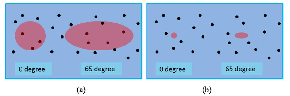 A further surface contamination or aggregation of mini-plasma may also affect the LIDT results. To verify whether these factors play a role, we put the 2 nm gold particles either on the surface or within the interface, as show in Fig. 8. Then, the laser damage experiment was carried out both with small and large laser beam sizes. The LIDT results demonstrate that the relative value of the LIDTs did not change regardless of whether the gold particles were on the surface or interface, the LIDT of the 65 degree HR coating was still lower than 0 degree HR coating for the large laser beam. However, all these experiments fail to explain the abnormal dependence of LIDT on EFI for large laser beam. Figure 8.Schematic presentation of HR coating with embedded gold nanoparticles on (a) surface and (b) interface  Since both of the micro-sized nodules and the nano-sized absorbers cannot explain the contradictory LIDT results with different beam size, we need to rethink the dominant damage mechanism for NUV HR coatings. Although the present theory still cannot explain this abnormal phenomenon very well, this remains an interesting and fascinating question, which merits further exploration of the damage mechanisms for NUV HR coatings. Furthermore, more information and understanding about this field will help us to find a more useful way to increase the LIDT of the NUV HR coatings. 4.CONCLUSIONSIn this study, the nanosecond laser-induced damage of NIR and NUV HR coatings were investigated. For NIR HR coatings, the physical insight of EFI enhancement at nodules are discussed. Light focusing and light penetrating have been proven to be the main mechanisms for creating the intensified EFI in the nodules. The focusing characteristics of the nodule can be affected by nodular geometry and refractive index of the medium, and the focal length of the nodule can be determined via a simplified model. Consequently, the light penetrating behaviors can be estimated via the ADT of the nodule. We conclude that strong EFI enhancement is developed when a certain portion of the light penetrates to the focal position of the nodules. For NUV HR coatings, our method that reduced the EFI within the coatings by increasing incident angle, showed totally different results for small and big testing beam sizes. Compared to 0 degree HR coating, 65 degree HR coating results in a much lower EFI. Its LIDT was higher for small testing beam size, but lower for large beam size. Several experiments were carried out to prove that both the micro-sized nodules and the nano-sized absorbers cannot explain this abnormal dependence of LIDT on EFI. We suspect, that some other mechanisms, such as plasma-material coupling, may play an important role in this phenomenon. More work is still required to further our understanding about this phenomenon. This work is partly supported by the National Natural Science Foundation of China (61522506, 51475335, 62621001, 61235011), the National Program on Key Research Project (2016YFA0200900), the National Key Scientific Instrument and Equipment Development Project (2014YQ090709), and the National Major Research Equipment Development project (ZDYZ2013-2). REFERENCESS. Juodkazis, K. Nishimura, S. Tanaka, H. Misawa, E. Gamaly, B. Luther-Davies, L. Hallo, P. Nicolai, and V. Tikhonchuk,
“Laser-induced microexplosion confined in the bulk of a sapphire crystal: Evidence of multi-megabar pressures,”
Phys. Rev. Lett., 96
(166101),
(2006). Google Scholar
B. C. Stuart, M. D. Feit, A. M. Rubenchik, B. W. Shore, and M. D. Perry,
“Laser-Induced Damage in Dielectrics with Nanosecond to Subpicosecond Pulses,”
Phys. Rev. Lett., 74 2248
–2251
(1995). https://doi.org/10.1103/PhysRevLett.74.2248 Google Scholar
J. Tauer, H. Kofler and E. Wintner,
“Laser-initiated ignition,”
Laser Photon. Rev., 4 99
–22
(2010). https://doi.org/10.1002/lpor.v4:1 Google Scholar
I. Kilen, J. Hader, J. V. Moloney, S. W. Koch,
“Ultrafast nonequilibrium carrier dynamics in semiconductor laser mode locking,”
Optica., 1 192
–197
(2014). https://doi.org/10.1364/OPTICA.1.000192 Google Scholar
A. A. Manenkov,
“Fundamental mechanisms of laser-induced damage in optical materials: today's state of understanding and problems,”
Opt. Eng., 53
(010901),
(2014). Google Scholar
L. Jensen, M. Mende, S. Schrameyer, M. Jupé, D. Ristau,
“Role of two-photon absorption in Ta2O5 thin films in nanosecond laser-induced damage,”
Opt. Lett., 37 4329
–4331
(2012). https://doi.org/10.1364/OL.37.004329 Google Scholar
Carr C W, Radousky H B, Rubenchik A M,
“Localized dynamics during laser-induced damage in optical material,”
Physical review letters, 92
(2004). https://doi.org/10.1103/PhysRevLett.92.087401 Google Scholar
Papernov S, Schmid A W.,
“Correlations between embedded single gold nanoparticles in SiO2 thin film and nanoscale crater formation induced by pulsed-laser radiation,”
Journal of Applied Physics, 92
(10), 5720
–5728
(2002). https://doi.org/10.1063/1.1512691 Google Scholar
Zhang D, Wang C, Fan P,
“Influence of plasma treatment on laser-induced damage characters of HfO 2 thin films at 355nm,”
Optics express, 17
(10), 8246
–8252
(2009). https://doi.org/10.1364/OE.17.008246 Google Scholar
Gallais L, Capoulade J, Natoli J Y,
“Investigation of nanodefect properties in optical coatings by coupling measured and simulated laser damage statistics,”
Journal of Applied Physics, 104
(5), 053120
(2008). https://doi.org/10.1063/1.2975179 Google Scholar
Carr C W, Radousky H B, Demos S G.,
“Wavelength dependence of laser-induced damage: determining the damage initiation mechanisms,”
Physical Review Letters, 91
(12), 127402
(2003). https://doi.org/10.1103/PhysRevLett.91.127402 Google Scholar
Dijon J, Poulingue M, Hue J.,
“Thermomechanical model of mirror laser damage at 1.06 μm I. Nodule ejection,”
Laser-Induced Damage in Optical Materials: 1998, 387
–397 International Society for Optics and Photonics,1999). Google Scholar
Dijon J, Ravel G, Andre B.,
“Thermomechanical model of mirror laser damage at 1.06 μm: II. Flat bottom pits formation,”
Laser-Induced Damage in Optical Materials: 1998, 398
–407 International Society for Optics and Photonics,1999). Google Scholar
Weakley S C, Stolz C J, Wu Z,
“Role of starting material composition in interfacial damage morphology of hafnia-silica multilayer coatings,”
Laser-Induced Damage in Optical Materials: 1998, 137
–143 International Society for Optics and Photonics,1999). Google Scholar
Guenther K H,
“Nodular defects in dielectric multilayers and thick single layers,”
Applied optics, 20
(6), 1034
–1038
(1981). https://doi.org/10.1364/AO.20.001034 Google Scholar
Cheng X, Shen Z, Jiao H,
“HfO2/SiO2 High Reflectors for 1.064 μm High Power Laser Applications,”
Optical Interference Coatings. Optical Society of America, FA9,
(2010). https://doi.org/10.1364/OIC.2010.FA9 Google Scholar
Cheng X, Ding T, He W,
“Using engineered defects to study laser-induced damage in optical thin films with nanosecond pulses,”
in XLIII Annual Symposium on Optical Materials for High Power Lasers. International Society for Optics and Photonics,
819002
–819002-9
(2011). Google Scholar
Cheng X, Ding T, He W,
“Using monodisperse SiO2 microspheres to study laser-induced damage of nodules in HfO2/SiO2 high reflectors,”
SPIE Optical Systems Design, 816816
–816816-6 International Society for Optics and Photonics,2011). https://doi.org/10.1117/12.896733 Google Scholar
Stolz C J, Feit M D, Pistor T V,
“Laser intensification by spherical inclusions embedded within multilayer coatings,”
Applied optics, 45
(7), 1594
–1601
(2006). https://doi.org/10.1364/AO.45.001594 Google Scholar
Stolz C J, Hafeman S, Pistor T V.,
“Light intensification modeling of coating inclusions irradiated at 351 and 1053 nm,”
Applied optics, 47
(13), C162
–C166
(2008). https://doi.org/10.1364/AO.47.00C162 Google Scholar
Milward J R, Lewis K L, Sheach K,
“Laser damage studies of silicon oxy-nitride narrowband reflectors,”
Optical Materials for High Power Lasers, 255
–264 International Society for Optics and Photonics,1993). https://doi.org/10.1117/12.147431 Google Scholar
Cheng X, Zhang J, Ding T,
“The effect of an electric field on the thermomechanical damage of nodular defects in dielectric multilayer coatings irradiated by nanosecond laser pulses,”
Light: Science & Applications, 2
(6),
(2013). https://doi.org/10.1038/lsa.2013.36 Google Scholar
Papernov S, Tait A, Bittle W,
“Near-ultraviolet absorption and nanosecond-pulse-laser damage in HfO2 monolayers studied by submicrometer-resolution photothermal heterodyne imaging and atomic force microscopy,”
Journal of Applied Physics, 109
(11),
(2011). https://doi.org/10.1063/1.3594713 Google Scholar
Papernov S, Schmid A W, Anzelotti J,
“AFM-mapped nanoscale absorber-driven laser damage in UV high-reflector multilayers,”
Laser-induced damage in optical materials: 1995, 384
–394 International Society for Optics and Photonics,1996). Google Scholar
Bonneau F, Combis P, Rullier J L,
“Study of UV laser interaction with gold nanoparticles embedded in silica,”
Applied Physics B, 75
(8), 803
–815
(2002). https://doi.org/10.1007/s00340-002-1049-7 Google Scholar
Papernov S, Schmid A W.,
“High-spatial-resolution studies of UV-laser-damage morphology in SiO2 thin films with artificial defects,”
in Boulder Damage Symposium XXXVI. International Society for Optics and Photonics,
141
–155
(2005). Google Scholar
Oliver J B, Papernov S, Schmid A W,
“Optimization of laser-damage resistance of evaporated hafnia films at 351nm,”
in Boulder Damage Symposium XL Annual Symposium on Optical Materials for High Power Lasers. International Society for Optics and Photonics,
71320J
–71320J-11
(2008). Google Scholar
Abromavicius G, Buzelis R, Drazdys R,
“Influence of electric field distribution on laser induced damage threshold and morphology of high reflectance optical coatings,”
in Boulder Damage Symposium XXXIX: Annual Symposium on Optical Materials for High Power Lasers. International Society for Optics and Photonics,
67200Y
–67200Y-8
(2007). Google Scholar
Xing H, Zhu M, Chai Y,
“Improving laser damage resistance of 355 nm high-reflective coatings by co-evaporated interfaces,”
Optics letters, 41
(6), 1253
–1256
(2016). https://doi.org/10.1364/OL.41.001253 Google Scholar
Cheng X, Tuniyazi A, Wei Z,
“Physical insight toward electric field enhancement at nodular defects in optical coatings,”
Optics express, 23
(7), 8609
–8619
(2015). https://doi.org/10.1364/OE.23.008609 Google Scholar
J.K. Murphy,
“Effects of surface and thin-film anomalies on miniature infrared filters,”
in Proc. SPIE,
64
–75
(1980). Google Scholar
J.R. Milward, K.L. Lewis, K. Sheach, and R. Heinecke,
“1.064 μm Laser Damage Studies of Silicon Oxy-Nitride Narrow Band Reflectors,”
in Proc. SPIE,
309
–316
(1994). Google Scholar
J. Dijon, M. Poulingue, and J. Hue,
“New approach for the critical size of the nodular defects: the mechanical connection,”
in Proc. SPIE,
370
–381
(1999). Google Scholar
C. J. Stolz, F. Y. Génina and T. V. Pistor,
“Electric-field enhancement by nodular defects in multilayer coatings irradiated at normal and 45° incidence,”
in Proc. SPIE,
41
–49
(2004). Google Scholar
Cheng X, Tuniyazi A, Zhang J,
“Nanosecond laser-induced damage of nodular defects in dielectric multilayer mirrors [Invited],”
Applied optics, 53
(4), A62
–A69
(2014). https://doi.org/10.1364/AO.53.000A62 Google Scholar
Dijon J, Poiroux T, Desrumaux C.,
“Nano absorbing centers: a key point in the laser damage of thin films,”
Laser-induced damage in optical materials: 1996, 315
–325 International Society for Optics and Photonics,1997). Google Scholar
Dijon J, Hue J, Disgecmez A,
“Thin film laser damage mechanisms at the YAG third harmonic,”
Laser-Induced Damage in Optical Materials: 1995, 416
–425 International Society for Optics and Photonics,1996). Google Scholar
Papernov S, Schmid A W.,
“Localized absorption effects during 351 nm, pulsed laser irradiation of dielectric multilayer thin films,”
Journal of applied physics, 82
(11), 5422
–5432
(1997). https://doi.org/10.1063/1.365570 Google Scholar
|


