|
|
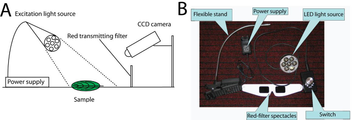
1.INTRODUCTIONIn 1931, H. Kautsky and A. Hirsch explored that the fluorescence emitted by the dark adapted plants is changing with the irradiation and its temporal variation is related to the photosynthesis1. Since it is an active effect which is influenced by several physiological and environmental interactions, it allows a deeper insight into the chemical and biological processes of the examined tissue compared to other, often claimed as passive, optical techniques (e.g. reflection or transmission). The chlorophyll (Chl) fluorescence of leaves consists of two maxima in the red (near 685–690 nm), and far-red region (near 730–740 nm). The intensity and shape of the Chl fluorescence emission spectrum of leaves at room temperature are dependent on the concentration of the fluorophore Chl-a, on the leaf structure, on the photosynthetic activity, and on the leaf’s optical properties2. However, the fluorescence induction kinetics (or transient) is caused by the change of the fluorescence yield with the onset of various processes of photosynthesis during the dark-to-light transfer3. Furthermore, a re-absorption is caused by the overlapping of the short wavelength range of the Chl fluorescence emission spectrum with the long-wavelength range of the Chl absorption spectrum, which can be used to estimate the active Chl content2. The aim of the paper is to present practical experiments, which introduce students into this biophysical, plant physiological research field. The laboratory practice is starting with a demonstration of the ‘Kautsky kinetic’ using an imaging setup. Then it continuous with a fluorescence kinetic measurement using a compact, intelligent fluorosensor (IFS), developed for basic and applied research on this field. 2.DEMONSTRATION WITH IMAGING EQUIPMENTIntact leaves, illuminated with strong light after a dark adaptation of at least 20 min, exhibit a transient change of fluorescence emission of the fluorophore Chl-a which is called ‘Kautsky effect’1. Time resolved investigation of the Chl-a fluorescence is widely used in basic and applied photosynthesis research3. For inducing photosynthetic activity, an actinic light is used which should be saturating, when measuring with continuous mode detection, or which should not be saturating when measuring with synchronous detection based on pulse amplitude modulation (PAM)3. Recently, PAM has become the most commonly used fluorescence induction technique for plant physiological studies and precision agriculture. However, we think that students need to see – likely with their own eyes – the basic fluorescence kinetic effect at first, in order to demonstrate them the basics of this technique and pique their interest for more complex methods and analyses. Therefore, we recommend start the laboratory practice with the demonstration of the ‘Kautsky effect’. For this purpose an intensive, e.g. LED, light source e.g. in the blue range is needed. Since the Chls are absorbing – of course with different efficiency – from the UV till ca. 700 nm4, other excitation wavelengths can be used as well. Any material for (partially) covering the leaf, and a red interference filter for detecting only the fluorescence wavebands and particularly excluding the excitation part of the spectrum, are needed. This filter can be used in form of a pair of red filter-spectacles and the students can observe the effect directly through these. The results described in this paper based on the demonstration kit developed by Opti-Sens Bt (e-mail: kocsanyi@eik.bme.hu). Or else, a capturing CCD camera can be equipped with a red filter and the images could be analyzed later on. As a result, a similar image sequence can be captured using a long (up to several seconds) exposition time which is showed on Figure 1. Perhaps, the two detections (one with human eye and other with CCD camera) can be realized simultaneously, if a bigger red-filter panel applied and students simultaneously with the CCD camera can observe the effect. We often use the leaves of Ficus benjamina L. or Laurus nobilis L. Necessarily we need a sample, which can be a living plant leaf or even an attached leaf, in order to simplify the setup. The experimental setup with a bigger red-filter panel shown on Figure 2 panel A, and the basic components of the setup using red filter-spectacles are shown on a photo on Figure 2 panel B. After this experiment with a living, functioning leaf, a fluorescence of a desiccated leaf need to be shown, which usually fluoresces at lower level and has almost no fluorescence kinetic. Figure 1.Captured images of the demonstration. The detected fluorescence image of partially dark adapted leaves of a Laurus nobilis L. plant A) immediately after finishing the dark adaptation and starting with the illumination B) after ca. 15 seconds illumination C) after ca. 90 seconds of illumination.  3.MEASUREMENT OF FLUORESCENCE PARAMETERS USING THE INTELLIGENT FLUOROSENSORAfter the students observed the basic process, we introduce them into more complex methods and teach them further biophysical phenomena. Usually, the fluorescence of leaves is monitored at dark adapted and light adapted states, and the photosynthesis is characterized by comparing the measured signals. The different adaptation states and the transition between them can be monitored by two different techniques: DC (continuous), total fluorescence measurement or PAM (pulse amplitude modulated), variable fluorescence signal. The total fluorescence emitted is always proportional to the excitation intensity which changes by several orders of magnitude. In case of the variable fluorescence, however, the measuring probe light, and the ‘saturating’ light and/or the so called actinic lights are separated. The last two determine the examinable light environment, whereas the measuring light provides the part of the excitation to which the response will be detected. As a result, only that portion of Chl-a fluorescence which is excited by an extremely low dose (low intensity and duty factor or on/off ratio) pulsed light of constant intensity termed as the probe light is measured5. The IFS provides total and variable fluorescence measurements simultaneously6. This has the advantage that the total fluorescence measurement – measured at two different wavebands at the two maxima of the Chl fluorescence – is based on the ‘Kautsky effect’, and the total fluorescence nicely shows the increasing photosynthetic activity and the simultaneously decreasing fluorescence signal. Therefore, these two fluorescence curves are easily understandable qualitatively after the demonstration experiment above. Since, in practice, we also apply so called saturating flashes, students see that the responses to the flashes also depend on the light adaptation of the photosynthetic reaction centers. From this, one can deduce the quenching analysis based on PAM technique with different (measuring, actinic and saturating) excitation light sources. Here we note that the final aim of the quenching analysis, based on the PAM technique, is to define the so-called photosynthetic energy partitioning, i.e. to define parameters (quantum yields) which will reflect the fraction of absorbed light energy utilized by particular processes (photochemistry vs. non-photochemical dissipations)7. Some important parameters are listed in Table 1. Table 1.Some examples for parameters which can be calculated from the fluorescence induction kinetic measurements. More parameters are listed in several publications3,7. PSI and PSII denote the two photosystems of the functioning photosynthetic apparatus operating in series3.
The measurement protocol of the IFS is completed by the leaf surface temperature measurement6,8. The core of the measurement protocol is shown in Figure 3 in case of a living leaf with functioning photosynthetic apparatus. The detailed measurement protocol is described by Barócsi6. Additionally, the fluorescence of a fully desiccated leaf should also be presented, which is only changing stepwise between different levels indicating that no more biophysical processes exist in a fully desiccated leaf. Figure 3.The core of the measurement protocol of the IFS. Letters mark the different excitations: (a) measuring probe light, (b): saturating flas, (c) actinic light and (d) far-red light  The IFS instrumentation is described in detail by Barócsi6, and the concept of the instrumentation is shown in Figure 4. The IFS tool is also capable to record the excitation kinetics9-10. This kind of measurement is based on a feedback mechanism. When keeping the fluorescence constant by the feedback mechanism of the IFS, the change of fluorescence yield during the dark-to-light transfer is compensated by an inverse change of the excitation light. The feedback mechanism reacts to the fluorescence changes of the sample and adjusts the laser power individually to the sample. Another feedback regulation adjusting the intensity of the excitation light in order to keep the Chl fluorescence stable is used by an instrument with a dual wavelength excitation (DUALEX)11. This technique is developed to acquire rapid information about the absorption characteristic of the leaf tissue and thus is used to ‘fully eliminate any artifacts caused by variable Chl fluorescence’ as has been demonstrated for quality assessment in grape vine production12. Therefore, it is also important to show this kind of measurement protocol for students, which has a clear potential in precision agriculture. Furthermore, the implementation of such a measurement technique is interesting for students studying physics or engineering. 4.CONCLUSIONSThe fluorescence induction kinetics of leaves is a complex signal, which depends on the concentration of the fluorophore Chl-a, on the leaf structure, on the photosynthetic activity and on the leaf’s optical properties. Here, we propose a progressive introduction of students – with the presumption having no preliminary experience – into this multidisciplinary research field. We start with the demonstration of the ‘Kautsky effect’, which shows the varying fluorescence caused by the change of fluorescence yield to photosynthetic processes during the dark-to-light transfer. Afterwards, more complex fluorescence studies are applied using IFS. The PAM measurement protocol allows the positioning of the photosynthetic energy using quenching analysis. Finally, the measurement protocol with feedback regulation is demonstrated. The proposed laboratory practice can be a good starting point for students interested for this multidisciplinary research field. ACKNOWLEDGEMENTSThe project was partially funded by,, TÁMOP-4.1.1.C-12/1/KONV-2012-0005 – Preparation of the concerned sectors for educational and R&D activities related to the Hungarian ELI project” supported by the European Union and co-financed by the European Social Fund. REFERENCESKautsky, H. and Hirsch, A.,
“Neue Versuche zur Kohlenstoffassimilation,”
Naturwissenschaften, 19 964
(1931). https://doi.org/10.1007/BF01516164 Google Scholar
Buschmann, C.,
“Variability and application of the chlorophyll fluorescence emission ratio red/far-red of leaves,”
Photosynth Res, 92 261
–271
(2007). https://doi.org/10.1007/s11120-007-9187-8 Google Scholar
Papageorgiou, G.C., Govindjee,
“Chlorophyll a Fluorescence, a Signature of Photosynthesis,”
Springer, Dordrecht
(2004). Google Scholar
Buschmann C., Lenk, S., Lichtenthaler, H. K.,
“Reflectance spectra and images of green leaves with different tissue structure and chlorophyll content,”
Israel Journal of Plant Sciences, 60
(1-2), 49
–64
(2012). https://doi.org/10.1560/IJPS.60.1-2.49 Google Scholar
Hout, Y., Babin M.,
“Overview of fluorescence protocols: theory, basic concepts and practiceChlorophyll a Fluorescence in Aquatic Sciences: Methods and Applications,”
Developments in Applied Phycology, 31
–74 Springer2011). Google Scholar
Barócsi, A.,
“Intelligent, net or wireless enabled fluorosensors for mass monitoring of assorted crops,”
Measurement Science and Technology, 24
(2), 1
–14
(2013). https://doi.org/10.1088/0957-0233/24/2/025701 Google Scholar
Lazár D.,
“Parameters of photosynthetic energy partitioning,”
Journal of Plant Physiology, 175 131
–147
(2015). https://doi.org/10.1016/j.jplph.2014.10.021 Google Scholar
Solti, Á., Lenk, S., Mihailova, G., Mayer, P., Barócsi, A., Georgieva, K.,
“Effects of habitat light conditions on the excitation quenching pathways in desiccating Haberlea rhodopensis leaves: An Intelligent FluoroSensor study,”
Journal of Photochemistry and Photobiology B: Biology, 130 217
–225
(2014). https://doi.org/10.1016/j.jphotobiol.2013.11.016 Google Scholar
Barocsi, A., Lenk, S., Kocsányi, L., Buschmann, C.,
“Excitation kinetics during induction of chlorophyll a fluorescence,”
Photosynthetica, 47
(1)), 104
–111
(2009). https://doi.org/10.1007/s11099-009-0016-5 Google Scholar
Buschmann, C., Konanz, S., Zhou, M., Lenk, S., Kocsányi, L., Barócsi, A.,
“Excitation kinetics of chlorophyll fluorescence during light-induced greening and establishment of photosynthetic activity of barley seedlings,”
Photosynthetica, 51
(2), 221
–230
(2013). https://doi.org/10.1007/s11099-013-0017-2 Google Scholar
Goulas, Y., Cerovic, Z.G., Cartelat, A., Moya, I.,
“Dualex: a new instrument for field measurements of epidermal ultraviolet absorbance by chlorophyll fluorescence,”
Applied Optics, 43 4488
–4496
(2004). https://doi.org/10.1364/AO.43.004488 Google Scholar
Cerovic, Z.G., Moise, N., Agati, G., Latouche, G., Ben Ghozlen, N., Meyer, S.,
“New portable optical sensors for the assessment of winegrape phenolic maturity based on berry fluorescence,”
Journal of Food Composition and Analysis, 21 650
–654
(2008). https://doi.org/10.1016/j.jfca.2008.03.012 Google Scholar
|